. 2012;7(1):e29438.
doi: 10.1371/journal.pone.0029438. Epub 2012 Jan 17.

Angiopoietin 2 alters pancreatic vascularization in diabetic conditions

Affiliations

Angiopoietin 2 alters pancreatic vascularization in diabetic conditions

Sophie Calderari et al. PLoS One. 2012.

Abstract

Aims/hypothesis: Islet vascularization, by controlling beta-cell mass expansion in response to increased insulin demand, is implicated in the progression to glucose intolerance and type 2 diabetes. We investigated how hyperglycaemia impairs expansion and differentiation of the growing pancreas. We have grafted xenogenic (avian) embryonic pancreas in severe combined immuno-deficient (SCID) mouse and analyzed endocrine and endothelial development in hyperglycaemic compared to normoglycaemic conditions.

Methods: 14 dpi chicken pancreases were grafted under the kidney capsule of normoglycaemic or hyperglycaemic, streptozotocin-induced, SCID mice and analyzed two weeks later. Vascularization was analyzed both quantitatively and qualitatively using either in situ hybridization with both mouse- and chick-specific RNA probes for VEGFR2 or immunohistochemistry with an antibody to nestin, a marker of endothelial cells that is specific for murine cells. To inhibit angiopoietin 2 (Ang2), SCID mice were treated with 4 mg/kg IP L1-10 twice/week.

Results: In normoglycaemic condition, chicken-derived endocrine and exocrine cells developed well and intragraft vessels were lined with mouse endothelial cells. When pancreases were grafted in hyperglycaemic mice, growth and differentiation of the graft were altered and we observed endothelial discontinuities, large blood-filled spaces. Vessel density was decreased. These major vascular anomalies were associated with strong over-expression of chick-Ang2. To explore the possibility that Ang2 over-expression could be a key step in vascular disorganization induced by hyperglycaemia, we treated mice with L1-10, an Ang-2 specific inhibitor. Inhibition of Ang2 improved vascularization and beta-cell density.

Conclusions: This work highlighted an important role of Ang2 in pancreatic vascular defects induced by hyperglycaemia.


Conflict of interest statement

Competing Interests: The authors have read the journal's policy and have the following conflicts: SC obtained a commercial source from SERVIER laboratories. This does not alter the authors' adherence to all the PLoS ONE policies on sharing data and materials.

Figures

Figure 1
Figure 1. Blood glucose concentrations of SCID mice during the 2 weeks post-graft of chicken pancreas.
Continious line, white square: control mice, n = 12. Continious line, black square: STZ mice, n = 7. Dotted line, white square: L1–10 treated control mice, n = 5. Dotted line, black square: L1–10 treated STZ mice, n = 5. Mice were used for grafting experiments between 3 to 5 days after STZ or citrate buffer injections. *** p<0.001 control and L1–10 treated control mice vs STZ and L1–10 treated STZ mice. $ p<0.05, $$$ p<0.001 control mice vs: L1–10 treated control mice.
Figure 2
Figure 2. Graft of embryonic chick pancreas under SCID mouse kidney capsule.
At 14 dpi embryonic chick pancreas (A) was grafted under the kidney capsule of a normoglycaemic SCID mouse and analyzed 2 weeks post-transplantation (B). At that time, hematoxylin-eosin coloration (C) enabled us to observe many vessels filled with erythrocytes (arrow). Immunohistochemistry for nestin (D) and in situ hybridization for mouse VEGFR2 probe (E) indicated the murine origin of endothelial cells in the pancreatic graft. In hyperglycaemic conditions (F–I), dark spots were present on the top of the pancreatic grafts (F). Vascularization was disorganized with large blood-fill spaces (G, arrow) associated with vessel discontinuities (H, insert). Morphometry analyses of erythrocytes staining (G), nestin immunohistochemistry (H) and mouse VEGFR2 in situ hybridization (I) showed a decreased number of endothelial cells (J, K and L). Student t-test *p<0.05, n = 4–6.
Figure 3
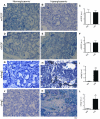
Figure 3. Endocrine and exocrine differentiation in normo and hyperglycaemic pancreas.
Insulin (A, D), glucagon (B, E) and amylase (C, F) positive-cells in 2 weeks grafted pancreata in normoglycaemic (A–C) and hyperglycaemic conditions (D–F). Quantification of insulin (G), glucagon (H) and amylase (I) positive-cells density. Hyperglycaemia increased beta-cell density (G). Student t- test *p<0.05, n = 3–5.
Figure 4
Figure 4. In situ hybridization of angiogenic factors in pancreatic grafts after 2 weeks hyperglycaemia.
Expression of mouse VEGF (A, B), chick VEGF (D, E), mouse Ang2 (G, H) and chick Ang2 (J, K) probes in normoglycemic (A, D, G, J) and hyperglycaemic grafts (B, E, H, K) was analyzed by in situ hybridization. C, F, I and L, semi-quantification of labeling intensity between normoglycaemic and hyperglycaemic conditions. Ang2 expressions were increased by hyperglycaemia. Student t-test, **p<0.01, ***p<0.001, n = 5.
Figure 5
Figure 5. Over-expression of VEGF using RCAS retrovirus.
Embryonic chick pancreas was infected with RCAS-GFP just before grafting. GFP was only and strongly expressed in the graft (A). In B, a photograph of a RCAS-VEGF-infected and grafted pancreas after 2 weeks hyperglycaemia showed the presence of blood filled spaces. In C, nestin immunohistochemsitry in VEGF-over-expressing and grafted pancreas after 2 weeks in hyperglycaemic conditions and in D, morphometry analyses of nestin immunohistochemistry (n = 4).
Figure 6
Figure 6. L1–10- induced specific Ang2 inhibition in hyperglycaemic conditions.
During the 2 weeks post-graft, STZ-induced diabetic SCID mice received L1–10 (4 mg/kg, twice-a-week). Pancreata were then collected (A) and vascularization was analyzed by hematoxylin-eosin staining (B) and nestin immunohistochemistry (C). Large blood-filled spaces were decreased by L1–10 treatment (E). Insulin staining (G) showed a trend towards increased β-cell density by L1–10 treatment.

References

    1. Eberhard D, Kragl M, Lammert E. ‘Giving and taking’: endothelial and beta-cells in the islets of Langerhans. Trends Endocrinol Metab. 2010;21:457–463. - PubMed
    1. Lammert E, Gu G, McLaughlin M, Brown D, Brekken R, et al. Role of VEGF-A in vascularization of pancreatic islets. Curr Biol. 2003;13:1070–1074. - PubMed
    1. Brissova M, Shostak A, Shiota M, Wiebe PO, Poffenberger G, et al. Pancreatic islet production of vascular endothelial growth factor–a is essential for islet vascularization, revascularization, and function. Diabetes. 2006;55:2974–2985. - PubMed
    1. Iwashita N, Uchida T, Choi JB, Azuma K, Ogihara T, et al. Impaired insulin secretion in vivo but enhanced insulin secretion from isolated islets in pancreatic beta cell-specific vascular endothelial growth factor-A knock-out mice. Diabetologia. 2007;50:380–389. - PubMed
    1. Richards OC, Raines SM, Attie AD. The role of blood vessels, endothelial cells, and vascular pericytes in insulin secretion and peripheral insulin action. Endocr Rev. 2010;31:343–363. - PMC - PubMed

Publication types

MeSH terms