. 2012;7(1):e29597.
doi: 10.1371/journal.pone.0029597. Epub 2012 Jan 17.

Capture of neuroepithelial-like stem cells from pluripotent stem cells provides a versatile system for in vitro production of human neurons

Affiliations

Capture of neuroepithelial-like stem cells from pluripotent stem cells provides a versatile system for in vitro production of human neurons

Anna Falk et al. PLoS One. 2012.

Abstract

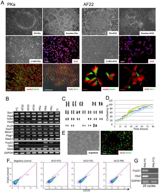
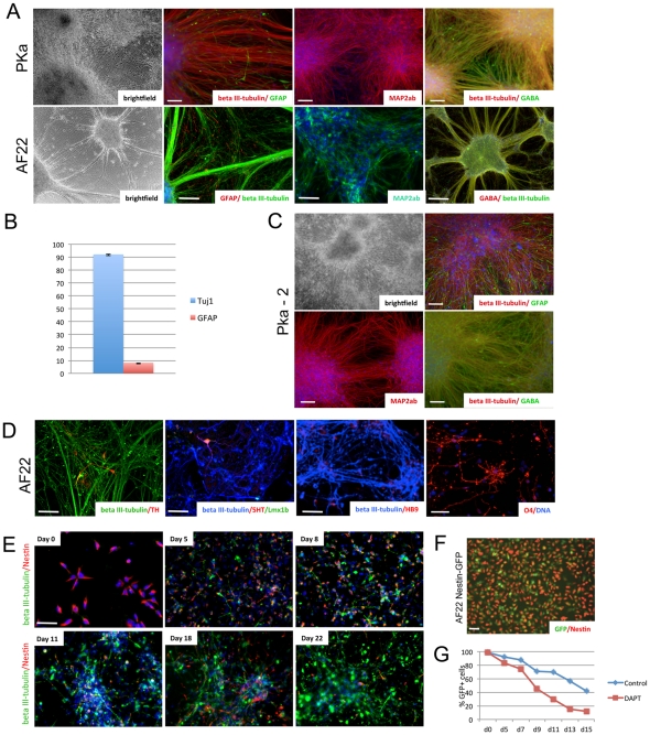
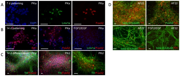
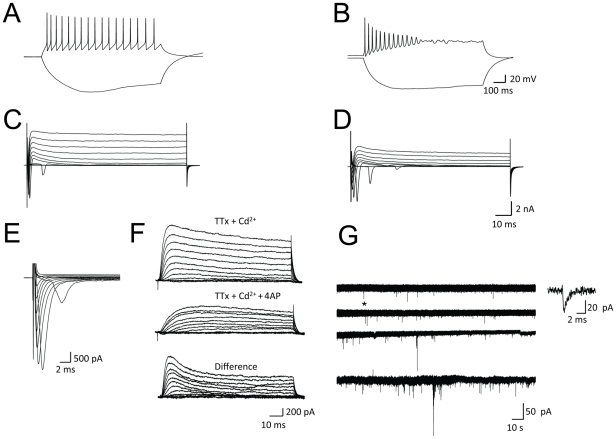
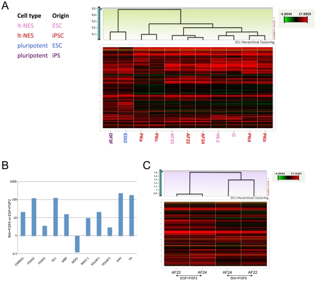
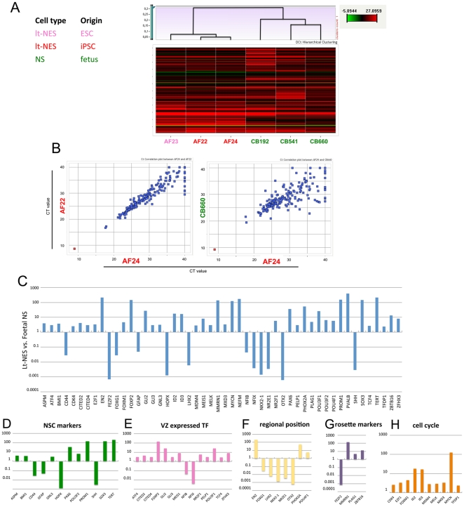
Human embryonic stem cells (hESC) and induced pluripotent stem cells (iPSC) provide new prospects for studying human neurodevelopment and modeling neurological disease. In particular, iPSC-derived neural cells permit a direct comparison of disease-relevant molecular pathways in neurons and glia derived from patients and healthy individuals. A prerequisite for such comparative studies are robust protocols that efficiently yield standardized populations of neural cell types. Here we show that long-term self-renewing neuroepithelial-like stem cells (lt-NES cells) derived from 3 hESC and 6 iPSC lines in two independent laboratories exhibit consistent characteristics including i) continuous expandability in the presence of FGF2 and EGF; ii) stable neuronal and glial differentiation competence; iii) characteristic transcription factor profile; iv) hindbrain specification amenable to regional patterning; v) capacity to generate functionally mature human neurons. We further show that lt-NES cells are developmentally distinct from fetal tissue-derived radial glia-like stem cells. We propose that lt-NES cells provide an interesting tool for studying human neurodevelopment and may serve as a standard system to facilitate comparative analyses of hESC and hiPSC-derived neural cells from control and diseased genetic backgrounds.


Conflict of interest statement

Competing Interests: OB is a co-founder of and has stock in LIFE & BRAIN GmbH. This does not alter the authors' adherence to all the PLoS ONE policies on sharing data and materials.

Figures

Figure 1
Figure 1. Long-term self-renewing neuroepithelial stem cells (lt-NES cells) derived from pluripotent stem cells of different origins.
(A) Representative pictures of the lt-NES cell lines PKa and AF22, derived from iPSCs generated from reprogrammed adult dermal fibroblasts. The iPSCs were induced to form neural rosettes, which were isolated and expanded into lt-NES cell lines. Lt-NES cells exhibit a rosette-like growth pattern, stain positive for Sox2, Dach1, Nestin and PLZF and show apical expression of the tight junction protein ZO-1. (B) Endpoint RT-PCR analysis (30 cycles) reveals expression of the neural progenitor markers SOX1, PAX6, HES5 and the neural rosette markers PLZF, DACH1, MMNR1 and PLAGL1. Lt-NES cells also express telomerase and high levels of the hindbrain marker GBX2. Fetal human cortex (FC) was used as control. (C) Lt-NES cells exhibit a stable karyotype over extensive passaging as assessed by G-banding (line AF22 at passage 30). (D) Growth kinetics of the three lt-NES cell lines AF22 (dark blue), AF23 (green), and AF24 (light blue) as measured by change in % confluence over time. (E) The clonal lt-NES line AF22:3, derived by single cell deposition into 96-well plates, displays a morphology indistinguishable from its parental line AF22 and stains positive for Sox2 and Nestin. (F) The cell surface expression of CD133/PROMININ by AF23 and AF22 lt-NES cells was analyzed using flow cytometry. Debris was excluded by use of TO-PRO-3, and both non-stained cells and cells stained with only the secondary antibody were used to set up the gate (negative control). (G) Early passage lt-NES cells (PKb, PKc) still express forebrain markers such as FOXG1 and OTX2. However, expression is lost at higher passages. P, passage number; white scale bars: 100 µm; red scale bars: 10 µm.
Figure 2
Figure 2. Neuronal differentiation of lt-NES cells.
(A–B) Four weeks after growth factor removal, lt-NES cells differentiated predominantly into neurons expressing beta III-tubulin (90%), MAP2ab and the neurotransmitter GABA, plus a minor fraction of GFAP-positive astrocytes (10%). Error bars represent STD. (C) Following growth factor withdrawal the clonal line PKa-2 gave rise to glia and neurons of preferentially GABAergic phenotype, comparable to the parental line PKa. (D) Specific neuronal subtypes expressing TH, 5HT and HB9 could be observed after 4 weeks of differentiation; O4-positive oligodendrocytes were detected after 10 weeks of differentiation. (E) AF22 lt-NES cells stained for Nestin and beta III-tubulin at day 0, 5, 8, 11, 18, 22 of differentiation. (F) Proliferating AF22 cells expressing GFP under the control of the Nestin enhancer (Nestin-GFP) show double labeling with an antibody to the Nestin protein. (G) Nestin-GFP expression in AF22 cells at day 0, 5, 7, 9, 11, 13, 15 of differentiation under control conditions or after exposure to DAPT (2 µM). Scale bars: 100 µm.
Figure 3
Figure 3. Induction of midbrain dopaminergic neurons using extrinsic factors.
IPS cell derived lt-NES cells cultured for 7 days in the presence of sonic hedgehog (Shh) and FGF8 showed nuclear expression of Lmx1a and FoxA2 (A). (B) With prolonged (14 days) exposure to Shh and FGF8, the number of cells expressing FoxA2 gradually increased. Shh/FGF8-treated cells were also positive for the midbrain marker En1, whereas neither FoxA2 nor Lmx1a were expressed in control populations cultured in the absence of Shh/FGF8. (C–D) Growth factor removal from lt-NES cell lines grown in Shh/FGF8 for 14 days induced differentiation and appearance of cell clusters positive for FoxA2 and tyrosine hydroxylase (TH), with beta III-tubulin-positive neurons co-expressing Nurr1. In contrast, control cultures propagated in FGF2/EGF remained TH-negative. Abbreviations in upper right corners of the immunofluorescence photomicrographs denote lt-NES cell lines used. Scale bars: 50 µm.
Figure 4
Figure 4. Lt-NES cells differentiate into functional neurons.
Representative traces of membrane potentials in differentiated PKa (A) and PKc (B) lt-NES cells upon depolarization by current injection. Voltage-dependent membrane currents measured from the same neurons (C–D). Depolarizing voltage steps elicited voltage-dependent inward and outward currents. The fast and transient inward current could be isolated by use of intracellular Cs+, extra- and intracellular TEA and extracellular Cd2+ (E). PKc-derived neurons also expressed 4-AP-sensitive outward currents (F). (G) Representative traces of spontaneous synaptic responses in PKa (two upper traces) and PKc (two lower traces) neurons cultured on a monolayer of mouse astrocytes. A single postsynaptic current recording (asterisk) is shown in larger magnification.
Figure 5
Figure 5. Lt-NES cell lines of various origins are similar to each other.
(A) Heat map and hierarchical cluster analysis of TLDA expression data from 11 cell lines comprising 2 pluripotent and 9 lt-NES cell lines of different origin. The hierarchical cluster analysis (Pearson) using complete linkage based on delta CT gene expression values reveals two major clusters representing the two different cell types visualized. (B) Genes significantly up-regulated (corrected p-value threshold 0.05) in Shh- and FGF8-treated lt-NES cell lines compared to control culture conditions (EGF and FGF2). (C) Cluster analysis displays higher similarity between Shh/FGF8-treated lt-NES cell lines than to the parental lt-NES cell line still cultured in EGF and FGF2.
Figure 6
Figure 6. Gene expression analysis of lt-NES cells and fetal NS cells.
(A) A total of six cell lines representing two cell types (lt-NES and fetal NS) of different origins were profiled using TLDAs. Hierarchical cluster visualization (Pearson) shows separation into two major clusters representing the two cell types analyzed. (B) Correlation plots based on CT gene expression values of the individual cell lines. The lt-NES cell lines AF24 and AF22 show a higher degree of correlation than the lt-NES cell line AF24 and the NS line CB660, although the latter carry the same genome (R = 0.95 vs. R = 0.76). Red dots represent the median aggregated CT expression value of the 18S endogenous control; blue dots indicate the median aggregated CT expression values of all other genes on the TLDA. (C) Relative levels of differently expressed genes (corrected p-value threshold 0.05) in 9 lt-NES cell lines and 3 fetal NS cell lines. (D–H) Panels depicting the expression of markers representing the categories neural stem cell (NSC) markers, ventricular zone (VZ) expressed transcription factors (TF), regional position, rosette genes, and cell cycle. Data in C–H are depicted at a Log10 scale with the mean expression in fetal NS cells set to 1. No significant differences in expression levels were detected in ESC- vs. iPSC-derived lt-NES cells.

References

    1. Takahashi K, Tanabe K, Ohnuki M, Narita M, Ichisaka T, et al. Induction of pluripotent stem cells from adult human fibroblasts by defined factors. Cell. 2007;131:861–872. - PubMed
    1. Takahashi K, Yamanaka S. Induction of pluripotent stem cells from mouse embryonic and adult fibroblast cultures by defined factors. Cell. 2006;126:663–676. - PubMed
    1. Koch P, Kokaia Z, Lindvall O, Brüstle O. Emerging concepts in neural stem cell research: autologous repair and cell-based disease modelling. Lancet Neurol. 2009;8:819–829. - PubMed
    1. Mattis VB, Svendsen CN. Induced pluripotent stem cells: a new revolution for clinical neurology? Lancet Neurol. 2011;10:383–394. - PubMed
    1. Hu BY, Weick JP, Yu J, Ma LX, Zhang XQ, et al. Neural differentiation of human induced pluripotent stem cells follows developmental principles but with variable potency. Proc Natl Acad Sci U S A. 2010;107:4335–4340. - PMC - PubMed

Publication types

MeSH terms

Substances