. 2012;7(1):e30095.
doi: 10.1371/journal.pone.0030095. Epub 2012 Jan 17.

T-cell responses to the DBLα-tag, a short semi-conserved region of the Plasmodium falciparum membrane erythrocyte protein 1

Affiliations

T-cell responses to the DBLα-tag, a short semi-conserved region of the Plasmodium falciparum membrane erythrocyte protein 1

Evelyn N Gitau et al. PLoS One. 2012.

Abstract

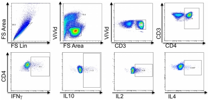
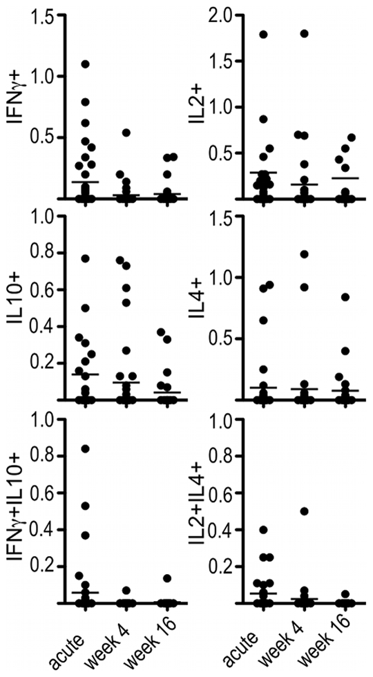
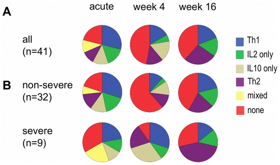
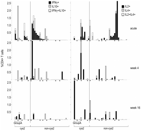
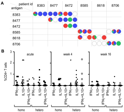
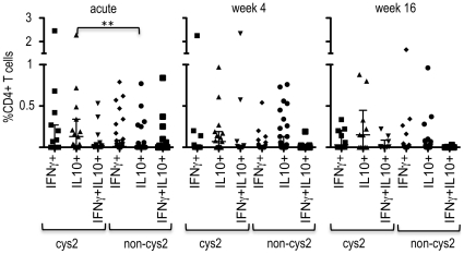
The Plasmodium falciparum erythrocyte membrane protein 1 (PfEMP1) is a variant surface antigen expressed on mature forms of infected erythrocytes. It is considered an important target of naturally acquired immunity. Despite its extreme sequence heterogeneity, variants of PfEMP1 can be stratified into distinct groups. Group A PfEMP1 have been independently associated with low host immunity and severe disease in several studies and are now of potential interest as vaccine candidates. Although antigen-specific antibodies are considered the main effector mechanism in immunity to malaria, the induction of efficient and long-lasting antibody responses requires CD4+ T-cell help. To date, very little is known about CD4+ T-cell responses to PfEMP1 expressed on clinical isolates. The DBLα-tag is a small region from the DBLα-domain of PfEMP1 that can be amplified with universal primers and is accessible in clinical parasite isolates. We identified the dominant expressed PfEMP1 in 41 individual clinical parasite isolates and expressed the corresponding DBLα-tag as recombinant antigen. Individual DBLα-tags were then used to activate CD4+ T-cells from acute and convalescent blood samples in children who were infected with the respective clinical parasite isolate. Here we show that CD4+ T-cell responses to the homologous DBLα-tag were induced in almost all children during acute malaria and maintained in some for 4 months. Children infected with parasites that dominantly expressed group A-like PfEMP1 were more likely to maintain antigen-specific IFNγ-producing CD4+ T-cells than children infected with parasites dominantly expressing other PfEMP1. These results suggest that group A-like PfEMP1 may induce long-lasting effector memory T-cells that might be able to provide rapid help to variant-specific B cells. Furthermore, a number of children induced CD4+ T-cell responses to heterologous DBLα-tags, suggesting that CD4+ T-cells may recognise shared epitopes between several DBLα-tags.


Conflict of interest statement

Competing Interests: The authors have declared that no competing interests exist.

Figures

Figure 1
Figure 1. Gating Strategy used to identify T-cell responses.
PBMCs were gated to eliminate doublets and dead. CD3+lymphocytes were identified and gated for CD4+ and CD8+ T-cells. Within each T-cell subset, the proportion of IFNγ, IL10, IL2, and IL4 producing cells were determined. IFNγ+IL10+ and IL2+IL4+ double producers were determined by Boolean gating.
Figure 2
Figure 2. Percentage of CD4+ T-cells producing cytokines after stimulation with the homologous DBLα-tag.
PBMCs from children obtained during acute malaria, 4 weeks and 16 weeks after the acute event were stimulated with recombinant DBLα-tag representing the dominant expressed PfEMP1 a given child was infected with. CD4+ T-cells were stained for production of INFγ and IL10 or IL2 and IL4 by intracellular cytokine staining. Shown are dot plots of percentages of CD4+ T-cells producing the indicated cytokine or cytokine combination during acute malaria or 4 and 16 weeks after the acute episode. Horizontal lines indicate the median.
Figure 3
Figure 3. Phenotype of CD4+ T-cells stimulated with the homologous DBLα-tag.
The phenotype of CD4+ T-cell in response to the homologous DBLα-tag was classified for each child and time point as followed: Th1, secretion of IFNγ alone or together with IL2 and/or IFNγ+IL10+; Th2, secretion of IL4 alone or in the presence of CD4+ T-cells secreting IL2 or IL10; IL2 alone: detection of only IL2-producing CD4+ T-cells; IL10 alone: detection of only IL10-producing CD4+ T-cells; mixed: detection of a mixed profile such as IFNγ-producing CD4+ T-cells together with IL4- or IL10-producing CD4+ T-cells or IL2- and IL10-producing CD4+ T-cells; none: children who did not induce any antigen-specific CD4+ T-cells. Shown are pie charts of the proportion of all children (A) with a particular profile or (B) grouped by disease severity.
Figure 4
Figure 4. Cumulative percentage of cytokine producing CD4+ T-cells after stimulation with homologous DBLα-tag by PfEMP1 subgroup.
Shown are cumulative bar-graphs of CD4+ T-cells producing cytokines indicated for each child. Data are grouped by the PfEMP1 subclass of the homologous DBLα-tag at acute disease, week 4 and week 16 after the acute event. Children suffering from severe disease are indicated by a star. Note that the order of children is the same in each plot.
Figure 5
Figure 5. CD4+ T-cells responses response to homologous or heterologous DBLα-tags.
(A) Representative examples of CD4+ T-cell responses to homologous and heterologous DBLα-tags for 6 children. Pie charts indicate the IFNγ+ (red), IL10+ (blue) and IFNγ+IL10+ double producing (green) CD4 T-cells as a proportion of the total cytokine producing CD4+ T-cells. White circles indicate the absence of a response. Within each cells, the sequence of circles indicates the proportion of CD4+ T-cells at acute disease (left), and 4 weeks (middle) and 16 weeks (right) after the acute episode. (B) Dot plots of the percentage of CD4+ T-cell responses to homologous (homo) and heterologous (hetero) DBLα-tags during acute malaria or 4 and 16 weeks after the acute attack. Horizontal lines indicate the median.
Figure 6
Figure 6. CD4+ T-cells responses response to cys2 and non-cys2 DBLα-tags.
Dot plots of the percentage of CD4+ T-cell responses to cys2 and non-cys2 DBLα-tags whether or not they represent homologous or heterologous DBLα-tags during acute malaria or 4 and 16 weeks after the acute attack. Horizontal lines indicate the median.

Similar articles

Cited by

References

    1. Miller LH, Baruch DI, Marsh K, Doumbo OK. The pathogenic basis of malaria. Nature. 2002;415:673–679. - PubMed
    1. Kyes S, Horrocks P, Newbold C. Antigenic variation at the infected red cell surface in malaria. Annu Rev Microbiol. 2001;55:673–707. - PubMed
    1. Warimwe GM, Keane TM, Fegan G, Musyoki JN, Newton CR, et al. Plasmodium falciparum var gene expression is modified by host immunity. Proc Natl Acad Sci U S A 2009 - PMC - PubMed
    1. Bull PC, Lowe BS, Kortok M, Molyneux CS, Newbold CI, et al. Parasite antigens on the infected red cell surface are targets for naturally acquired immunity to malaria. Nat Med. 1998;4:358–360. - PMC - PubMed
    1. Bull PC, Lowe BS, Kortok M, Marsh K. Antibody recognition of Plasmodium falciparum erythrocyte surface antigens in Kenya: evidence for rare and prevalent variants. Infect Immun. 1999;67:733–739. - PMC - PubMed

Publication types

MeSH terms

Associated data