

On dorsal prothoracic appendages in treehoppers (Hemiptera: Membracidae) and the nature of morphological evidence
- PMID: 22272287
- PMCID: PMC3260216
- DOI: 10.1371/journal.pone.0030137
On dorsal prothoracic appendages in treehoppers (Hemiptera: Membracidae) and the nature of morphological evidence
Abstract
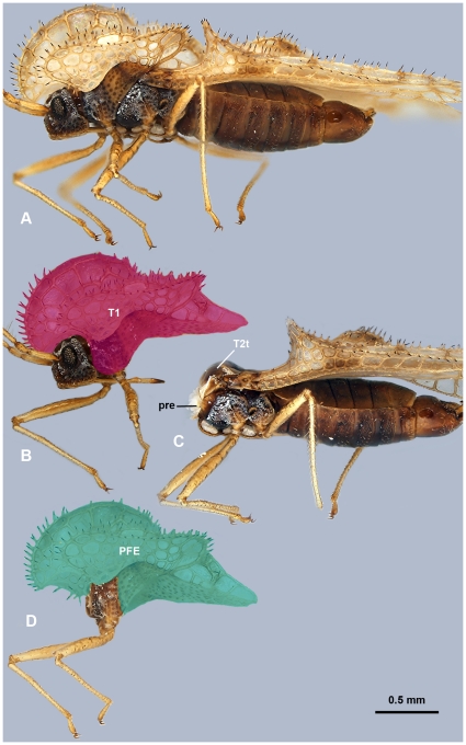
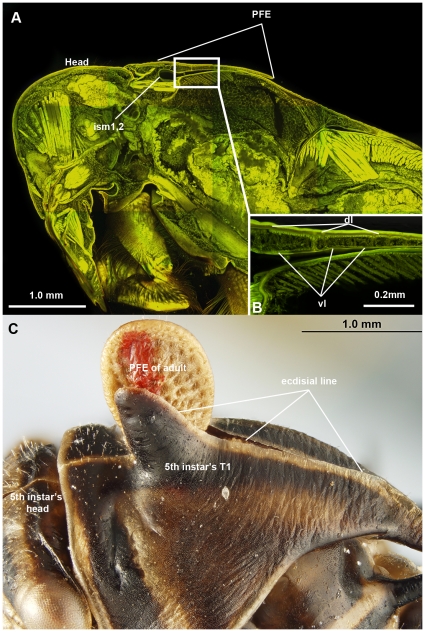
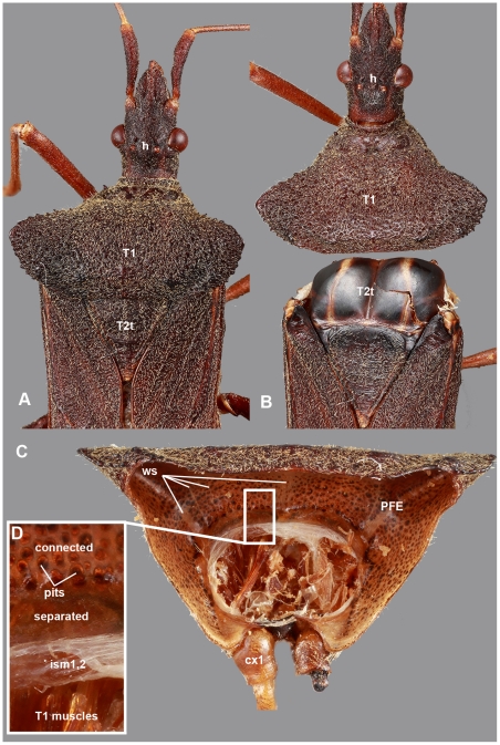
A spectacular hypothesis was published recently, which suggested that the "helmet" (a dorsal thoracic sclerite that obscures most of the body) of treehoppers (Insecta: Hemiptera: Membracidae) is connected to the 1st thoracic segment (T1; prothorax) via a jointed articulation and therefore was a true appendage. Furthermore, the "helmet" was interpreted to share multiple characteristics with wings, which in extant pterygote insects are present only on the 2nd (T2) and 3rd (T3) thoracic segments. In this context, the "helmet" could be considered an evolutionary novelty. Although multiple lines of morphological evidence putatively supported the "helmet"-wing homology, the relationship of the "helmet" to other thoracic sclerites and muscles remained unclear. Our observations of exemplar thoraces of 10 hemipteran families reveal multiple misinterpretations relevant to the "helmet"-wing homology hypothesis as originally conceived: 1) the "helmet" actually represents T1 (excluding the fore legs); 2) the "T1 tergum" is actually the anterior dorsal area of T2; 3) the putative articulation between the "helmet" and T1 is actually the articulation between T1 and T2. We conclude that there is no dorsal, articulated appendage on the membracid T1. Although the posterior, flattened, cuticular evagination (PFE) of the membracid T1 does share structural and genetic attributes with wings, the PFE is actually widely distributed across Hemiptera. Hence, the presence of this structure in Membracidae is not an evolutionary novelty for this clade. We discuss this new interpretation of the membracid T1 and the challenges of interpreting and representing morphological data more broadly. We acknowledge that the lack of data standards for morphology is a contributing factor to misinterpreted results and offer an example for how one can reduce ambiguity in morphology by referencing anatomical concepts in published ontologies.
Conflict of interest statement
Figures


Similar articles
-
Body plan innovation in treehoppers through the evolution of an extra wing-like appendage.Nature. 2011 May 5;473(7345):83-6. doi: 10.1038/nature09977. Nature. 2011. PMID: 21544145
-
Co-option of wing-patterning genes underlies the evolution of the treehopper helmet.Nat Ecol Evol. 2020 Feb;4(2):250-260. doi: 10.1038/s41559-019-1054-4. Epub 2019 Dec 9. Nat Ecol Evol. 2020. PMID: 31819237
-
Jumping mechanisms of treehopper insects (Hemiptera, Auchenorrhyncha, Membracidae).J Exp Biol. 2013 Mar 1;216(Pt 5):788-99. doi: 10.1242/jeb.078741. Epub 2012 Nov 15. J Exp Biol. 2013. PMID: 23155084
-
What crustaceans can tell us about the evolution of insect wings and other morphologically novel structures.Curr Opin Genet Dev. 2021 Aug;69:48-55. doi: 10.1016/j.gde.2021.02.008. Epub 2021 Feb 26. Curr Opin Genet Dev. 2021. PMID: 33647834 Review.
-
The evolution of insect wings and their sensory apparatus.Brain Behav Evol. 1997 Jul;50(1):13-24. doi: 10.1159/000113318. Brain Behav Evol. 1997. PMID: 9209763 Review.
Cited by
-
Evolution of thorax architecture in ant castes highlights trade-off between flight and ground behaviors.Elife. 2014;3:e01539. doi: 10.7554/eLife.01539. Epub 2014 Jan 7. Elife. 2014. PMID: 24399458 Free PMC article.
-
Developmental Transcriptomics Reveals a Gene Network Driving Mimetic Color Variation in a Bumble Bee.Genome Biol Evol. 2021 Jun 8;13(6):evab080. doi: 10.1093/gbe/evab080. Genome Biol Evol. 2021. PMID: 33881508 Free PMC article.
-
Structure and development of the complex helmet of treehoppers (Insecta: Hemiptera: Membracidae).Zoological Lett. 2020 Feb 24;6:3. doi: 10.1186/s40851-020-00155-7. eCollection 2020. Zoological Lett. 2020. PMID: 32123574 Free PMC article.
-
Insights into insect wing origin provided by functional analysis of vestigial in the red flour beetle, Tribolium castaneum.Proc Natl Acad Sci U S A. 2013 Oct 15;110(42):16951-6. doi: 10.1073/pnas.1304332110. Epub 2013 Oct 1. Proc Natl Acad Sci U S A. 2013. PMID: 24085843 Free PMC article.
-
What serial homologs can tell us about the origin of insect wings.F1000Res. 2017 Mar 14;6:268. doi: 10.12688/f1000research.10285.1. eCollection 2017. F1000Res. 2017. PMID: 28357056 Free PMC article. Review.
References
-
- Prud'homme B, Minervino C, Hocine M, Cande JD, Aouane A, et al. Body plan innovation in treehoppers through the evolution of an extra wing-like appendage. Nature. 2011;473:83–86. doi: 10.1038/nature09977. - DOI - PubMed
-
- Moczek AP. Evolutionary biology: The origins of novelty. Nature, 2011;473:34–35. - PubMed
-
- Pennisi E. Treehopper camouflage derives from ancestral wing. 2011. Available: http://news.sciencemag.org/sciencenow/2011/05/treehopper-camouflage-deri... Accessed 5 August 2011.
-
- Yong E. From 250 million years of repression, a wonderland of hats. 2011 Available: http://blogs.discovermagazine.com/notrocketscience/2011/05/04/from-250-m... Accessed 2011 August 5.
-
- Li H, Popadić A. Analysis of nubbin expression patterns in insects. Evol Dev. 2004;6:310–324. - PubMed
Publication types
MeSH terms
LinkOut - more resources
Full Text Sources
Other Literature Sources
