. 2012;7(1):e30294.
doi: 10.1371/journal.pone.0030294. Epub 2012 Jan 17.

Wogonin improves histological and functional outcomes, and reduces activation of TLR4/NF-κB signaling after experimental traumatic brain injury

Affiliations

Wogonin improves histological and functional outcomes, and reduces activation of TLR4/NF-κB signaling after experimental traumatic brain injury

Chien-Cheng Chen et al. PLoS One. 2012.

Erratum in

Abstract

Background: Traumatic brain injury (TBI) initiates a neuroinflammatory cascade that contributes to neuronal damage and behavioral impairment. This study was undertaken to investigate the effects of wogonin, a flavonoid with potent anti-inflammatory properties, on functional and histological outcomes, brain edema, and toll-like receptor 4 (TLR4)- and nuclear factor kappa B (NF-κB)-related signaling pathways in mice following TBI.

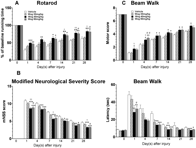
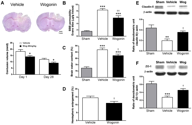
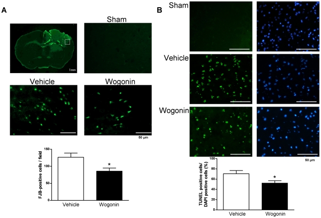
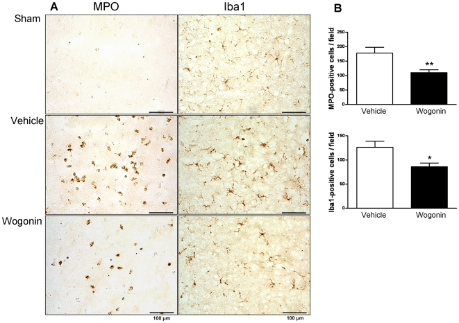
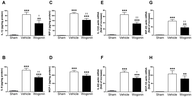
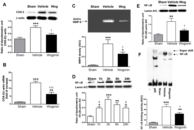
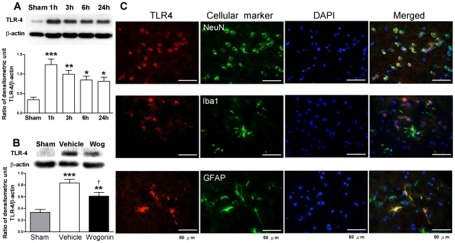
Methodology/principal findings: Mice subjected to controlled cortical impact injury were injected with wogonin (20, 40, or 50 mg·kg(-1)) or vehicle 10 min after injury. Behavioral studies, histology analysis, and measurement of blood-brain barrier (BBB) permeability and brain water content were carried out to assess the effects of wogonin. Levels of TLR4/NF-κB-related inflammatory mediators were also examined. Treatment with 40 mg·kg(-1) wogonin significantly improved functional recovery and reduced contusion volumes up to post-injury day 28. Wogonin also significantly reduced neuronal death, BBB permeability, and brain edema beginning at day 1. These changes were associated with a marked reduction in leukocyte infiltration, microglial activation, TLR4 expression, NF-κB translocation to nucleus and its DNA binding activity, matrix metalloproteinase-9 activity, and expression of inflammatory mediators, including interleukin-1β, interleukin-6, macrophage inflammatory protein-2, and cyclooxygenase-2.

Conclusions/significance: Our results show that post-injury wogonin treatment improved long-term functional and histological outcomes, reduced brain edema, and attenuated the TLR4/NF-κB-mediated inflammatory response in mouse TBI. The neuroprotective effects of wogonin may be related to modulation of the TLR4/NF-κB signaling pathway.


Conflict of interest statement

Competing Interests: The authors have declared that no competing interests exist.

Figures

Figure 1
Figure 1. Effects of 3 different doses of wogonin on functional outcomes in contusion-injured mice.
(A) Compared to the vehicle treatment, 20 mg·kg−1 wogonin treatment did not significantly alter rotarod performance. On days 1–28, 40 mg·kg−1 wogonin-treated mice performed significantly better than the vehicle-treated mice. On days 1, 7, 14, 21, and 28, the 50 mg·kg−1 wogonin-treated mice had better rotarod performance than the vehicle-treated mice. No significant differences were observed between the 40 mg·kg−1 and 50 mg·kg−1 wogonin treatment groups at any time point. (B) No difference in modified Neurological Severity Score (mNSS) was detected between the 20 mg·kg−1 wogonin and vehicle treatment groups. On days 1–28, the mNSSs were significantly lower in the 40 mg·kg−1 and 50 mg·kg−1 dose groups than in the vehicle-treated group. No significant differences were observed between the 40 mg·kg−1 and 50 mg·kg−1 treatment groups. (C) During the beam walk test, no significant differences were observed between the 20 mg·kg−1 wogonin-treated and vehicle-treated groups. Significant differences were observed in the hindlimb motor scores between the 40 mg·kg−1 wogonin-treated and vehicle-treated groups on post-injury test days 1–28 and between the 50 mg·kg−1 dose group and vehicle group on days 4, 7, 14, and 21. Differences in hindlimb motor scores between the 40 mg·kg−1 and 50 mg·kg−1 groups were not significant. Beam walk latencies were significantly shorter for both the 40 mg·kg−1 and 50 mg·kg−1 groups than for the vehicle group, on days 1, 7, 14, and 28; however, no significant difference was observed between the 2 wogonin-treated groups. Values are presented as mean ± SEM; *P<0.05, **P<0.01, ***P<0.001 versus vehicle-treated injured mice. P<0.05, †† P<0.01: 20 mg·kg−1 wogonin-treated mice versus the 40 mg·kg−1 wogonin-treated mice. # P<0.05: 20 mg·kg−1 wogonin-treated mice versus the 50 mg·kg−1 wogonin-treated mice (n = 8 mice/group at each time point).
Figure 2
Figure 2. Effects of 40 mg·kg−1 wogonin treatment on cortical contusion volume, brain edema, and BBB permeability.
(A) Representative cresyl violet-stained brain sections of vehicle- and 40 mg·kg−1 wogonin-treated mice 1 day post-TBI showing hypointense regions immediately below the impact site in the cortex. Scale bar is 1 mm. Quantification showed significantly smaller contusion volumes in wogonin-treated mice compared with vehicle-treated mice at days 1 and 28 post-TBI. (B) Wogonin-treated mice showed a significant decrease in the concentration of Evans blue (EB) in the ipsilateral hemisphere compared with vehicle-treated mice at day 1. (C) Brain water content in the ipsilateral hemisphere of 40 mg·kg−1 wogonin-treated mice was significantly lower than in vehicle-treated mice at day 1. (D) At day 1, hemispheric enlargement was significantly smaller in mice treated with 40 mg·kg−1 wogonin than in vehicle-treated mice. (E, F) Treatment with 40 mg·kg−1 wogonin also reversed TBI-mediated reduced expression of claudin-5 and zonula occludens 1 in traumatic cortical areas of the ipsilateral hemisphere at day 1 following TBI, as measured by western blot. Values are presented as mean ± SEM; **P<0.01, ***P<0.001 versus vehicle-treated injured mice. P<0.05, †† P<0.01 for the 40 mg·kg−1 wogonin-treated mice versus vehicle-treated mice. (n = 8 mice/group for cresyl violet staining and hemispheric enlargement, n = 7 mice/group for brain EB, brain water content, and western blot).
Figure 3
Figure 3. Effects of 40 mg·kg−1 wogonin treatment on neuronal degeneration and apoptotic cell death.
(A) Brain atlas of coronal sections of a core contusional region at 0.74 mm from the bregma. Quantification analysis indicated that wogonin-treated mice had significantly fewer degenerating neurons than vehicle-treated mice in the cortical contusion margin at day 1 post-TBI. The total number of Fluoro-Jade B (FJB)-positive cells is expressed as the mean number per field of view (1.3 mm2). The scale bar is 50 µm. (B) Representative terminal deoxynucleotidyl transferase-mediated dUTP-biotin nick end labeling (TUNEL) staining (green)- and DAPI (blue)-stained brain sections of a sham-injured control, a wogonin-treated mouse, and a vehicle-treated mouse at day 1 post-TBI. Quantification showed that wogonin-treated mice had significantly fewer TUNEL-positive cells than the vehicle-treated mice in the cortical contusion margin at day 1 post-TBI. The percentage of TUNEL-positive cells is expressed as the number of TUNEL-stained nuclei/the total number of DAPI-stained nuclei. Sections were stained with DAPI (blue) to show all nuclei. The scale bar is 50 µm. Values are presented as means ± SEM; *P<0.05 versus vehicle-treated injured mice (n = 7 mice/group).
Figure 4
Figure 4. Effects of 40 mg·kg−1 wogonin treatment on neutrophil infiltration and microglial activation.
(A) Representative myeloperoxidase (MPO)- and anti-ionized calcium binding adaptor molecule 1 (Iba1)-stained brain sections from a sham-injured control, a wogonin-treated, and a vehicle-treated mouse at day 1 post-TBI. (B) Cell count analysis indicated that wogonin-treated mice had significantly fewer infiltrating neutrophils and activated microglia/macrophages than vehicle-treated mice in the cortical contusion margin at day 1 post-TBI. The total number of MPO- and Iba1-positive cells is expressed as the mean number per field of view (1.3 mm2). The scale bar is 100 µm. Values are presented as means ± SEM; *P<0.05, **P<0.01 versus vehicle-treated TBI mice (n = 7 mice/group).
Figure 5
Figure 5. Effects of 40 mg·kg−1 wogonin treatment on protein and mRNA expression of cytokines and chemokines.
(A, B, C, D) Bar graphs of interleukin (IL)-1β, IL-6, macrophage inflammatory protein (MIP)-2, and monocyte chemoattractant protein (MCP)-1 protein concentrations in the ipsilateral cortices of sham control, vehicle-treated, and 40 mg·kg−1 wogonin-treated mice at day 1 post-injury. Wogonin significantly attenuated injury-induced increases in IL-1β, IL-6, and MIP-2 protein concentrations, but had no effect on MCP-1 protein concentration compared with vehicle-treated TBI mice. (E, F, G, H) Bar graphs demonstrating IL-1β, IL-6, MIP-2, and MCP-1 mRNA expression in the ipsilateral cortices of sham control, vehicle-treated, and 40 mg·kg−1 wogonin-treated mice 6 h post-injury. Wogonin significantly inhibited injury-induced expression of IL-1β, IL-6, and MIP-2 mRNA in the ipsilateral cortices. There was no significant difference in MCP-1 mRNA transcript levels between the wogonin-treated and vehicle-treated groups of mice subjected to TBI. Values are presented as means ± SEM; **P<0.01, ***P<0.001 versus sham control, and P<0.05, †† P<0.01 for wogonin-treated mice versus vehicle-treated TBI mice (n = 7 mice/group).
Figure 6
Figure 6. Effects of 40 mg·kg−1 wogonin on COX-2 expression, MMP-9 and NF-κB activation.
(A) Representative immunoblots and densitometry analysis showed a significant decrease in COX-2 protein levels in the ipsilateral hemispheres of wogonin-treated injured mice at day 1 compared with vehicle-treated injured mice. (B) Bar graphs showed that wogonin significantly reduced COX-2 mRNA expression in the ipsilateral hemispheres compared with vehicle-treated injured mice at 6 h post-TBI. (C) Representative zymography and densitometry analysis showed that MMP-9 activity was significantly decreased in wogonin-treated mice compared with vehicle-treated mice at day 1. (D) Representative immunoblots of nuclearNF-κB p65 in the ipsilateral hemisphere of untreated injured mice. Densitometric analysis showed increased nuclear p65 levels at 1, 3, 6 and 24 h post-injury, with a peak at around 3 h. # P<0.05 versus 3 h (n = 5 mice/time point). (E) Representative immunoblots and densitometric analysis showed a significantly greater decrease in nuclear NF-κB p65 levels in the ipsilateral hemispheres of the wogonin-treated injured mice than in the vehicle-treated injured mice at day 1. (F) Representative gel shift analysis showing NF-κB DNA-binding activity from a sham control mouse (lane 3), vehicle-treated injured mouse (lane 4), and wogonin-treated injured mouse (lane 5) at day 1. Competition assays for NF-κB DNA-binding activity were performed with a 50-fold excess of unlabeled competitor NF-κB consensus oligonucleotides (lane 1). For the supershift assay, an antibody targeting the p65 subunit of NF-κB was incubated with a nuclear protein sample before the binding reaction (lane 2). Quantification analysis showed that wogonin treatment induced a significantly greater decrease in NF-κB binding activity, expressed in arbitrary densitometric units (ADU), than that induced by vehicle treatment, measured by EMSA.Values are reported as means ± SEM; *P<0.05, **P<0.01, ***P<0.001 versus sham controls, and P<0.05, ††† P<0.001 for wogonin-treated versus vehicle-treated TBI mice (n = 7 mice/group).
Figure 7
Figure 7. Effects of 40 mg·kg−1 wogonin on TLR4 expresssion.
(A) Representative immunoblots of toll-like receptor (TLR)-4 protein in the ipsilateral hemisphere from untreated injured mice. Bar graphs of densitometry analysis of the protein bands showed increased TLR4 levels at 1, 3, 6 and 24 h post-injury (n = 5 mice/time point). (B) Representative immunoblots showing TLR-4 protein in the ipsilateral hemisphere from a sham-injured control, a wogonin-treated injured mouse, and a vehicle-treated injured mouse at day 1 post-TBI. Bar graphs of densitometry analysis of protein bands showed a significant decrease in TLR4 protein levels in the ipsilateral hemispheres of wogonin-treated mice at day 1 post-TBI compared with vehicle-treated mice. (C) Identification of TLR4- positive cells 1 day post-injury in the peri-contusion margin by immunofluorescence labeling. TLR-4 immunoreactivity is shown in red, and immunolabeling of NeuN (a cell marker for neurons), anti-ionized calcium binding adaptor molecule 1 (Iba1, a cell marker for microglia), or GFAP (a cell marker for astrocytes) is shown in green. Yellow labeling indicates co-localization. TLR4 was co-localized in neurons and astrocytes, and poor colocolization was observed in microglia. Sections were stained with DAPI (blue) to show all nuclei. The scale bar is 50 µm. Values are reported as means ± SEM; *P<0.05, **P<0.01, ***P<0.001 versus sham controls, and P<0.05 for wogonin-treated versus vehicle-treated TBI mice (n = 7 mice/group).

Similar articles

Cited by

References

    1. Morganti-Kossmann MC, Rancan M, Stahel PF, Kossmann T. Inflammatory response in acute traumatic brain injury: a double-edged sword. Curr Opin Crit Care. 2002;8:101–105. - PubMed
    1. Downes CE, Crack PJ. Neural injury following stroke: are Toll-like receptors the link between the immune system and the CNS? Br J Pharmacol. 2010;160:1872–1888. - PMC - PubMed
    1. Dong XQ, Yu WH, Hu YY, Zhang ZY, Huang M. Oxymatrine reduces neuronal cell apoptosis by inhibiting Toll-like receptor 4/nuclear factor kappa-B-dependent inflammatory responses in traumatic rat brain injury. Inflamm Res. 2011;60:533–539. - PubMed
    1. Chen G, Shi J, Jin W, Wang L, Xie W, et al. Progesterone administration modulates TLRs/NF-kappaB signaling pathway in rat brain after cortical contusion. Ann Clin Lab Sci. 2008;38:65–74. - PubMed
    1. Chen G, Zhang S, Shi J, Ai J, Qi M, et al. Simvastatin reduces secondary brain injury caused by cortical contusion in rats: possible involvement of TLR4/NF-kappaB pathway. Exp Neurol. 2009;216:398–406. - PubMed

Publication types

MeSH terms