. 2012 Feb;9(1):20-31.
doi: 10.2174/156720212799297137.

Wnt1 inducible signaling pathway protein 1 (WISP1) blocks neurodegeneration through phosphoinositide 3 kinase/Akt1 and apoptotic mitochondrial signaling involving Bad, Bax, Bim, and Bcl-xL

Affiliations

Wnt1 inducible signaling pathway protein 1 (WISP1) blocks neurodegeneration through phosphoinositide 3 kinase/Akt1 and apoptotic mitochondrial signaling involving Bad, Bax, Bim, and Bcl-xL

Shaohui Wang et al. Curr Neurovasc Res. 2012 Feb.

Abstract

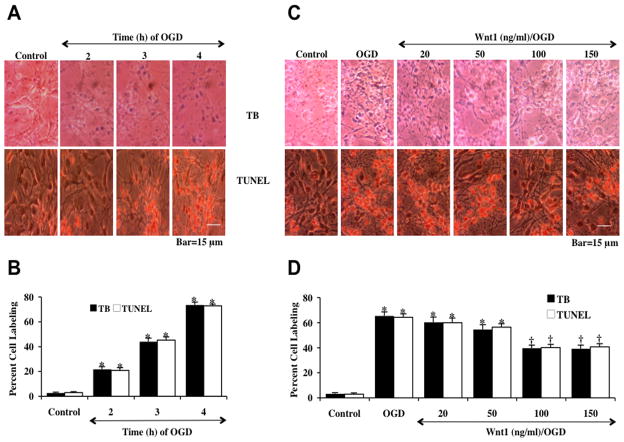
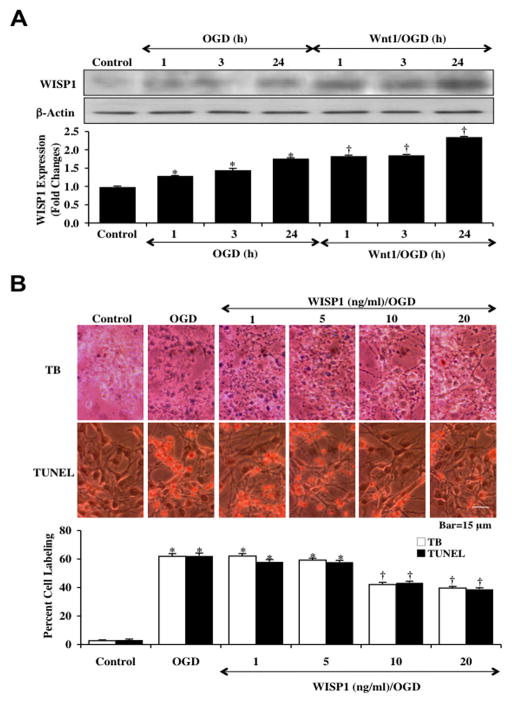
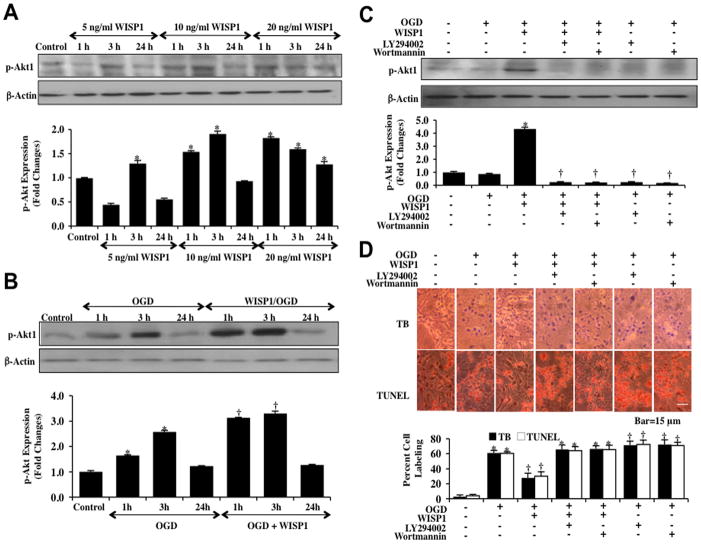
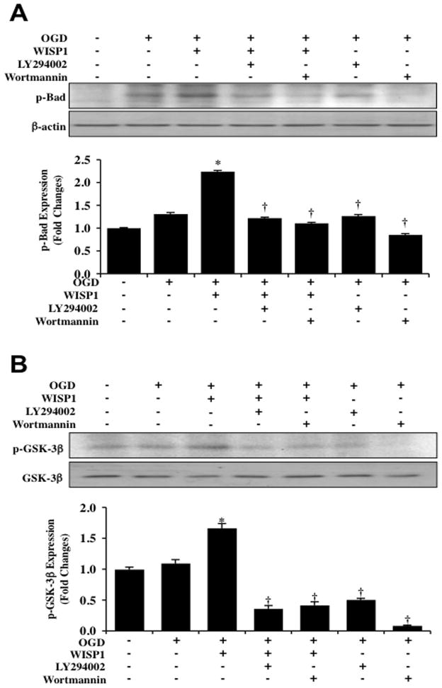
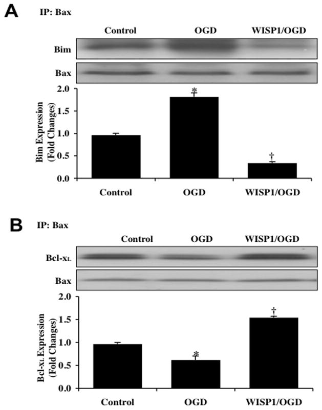
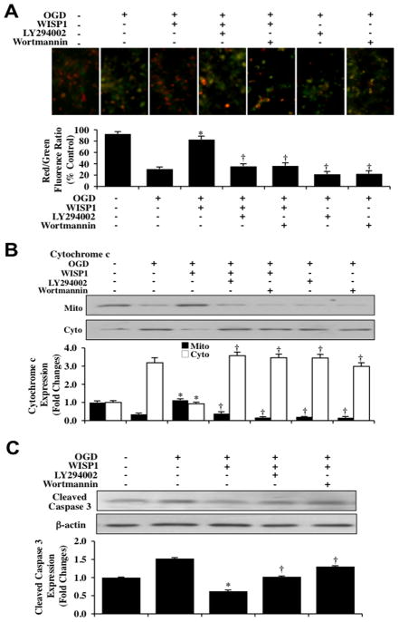
Wnt1 inducible signaling pathway protein 1 (WISP1) is a member of the CCN family of proteins that determine cell growth, cell differentiation, immune system activation, and cell survival in tissues ranging from the cardiovascular-pulmonary system to the reproductive system. Yet, little is known of the role of WISP1 as a neuroprotective entity in the nervous system. Here we demonstrate that WISP1 is present in primary hippocampal neurons during oxidant stress with oxygen-glucose deprivation (OGD). WISP1 expression is significantly enhanced during OGD exposure by the cysteine-rich glycosylated protein Wnt1. Similar to the neuroprotective capabilities known for Wnt1 and its signaling pathways, WISP1 averts neuronal cell injury and apoptotic degeneration during oxidative stress exposure. WISP1 requires activation of phosphoinositide 3-kinase (PI 3-K) and Akt1 pathways to promote neuronal cell survival, since blockade of these pathways abrogates cellular protection. Furthermore, WISP1 through PI 3-K and Akt1 phosphorylates Bad and GSK-3β, minimizes expression of the Bim/Bax complex while increasing the expression of Bclx(L)/Bax complex, and prevents mitochondrial membrane permeability, cytochrome c release, and caspase 3 activation in the presence of oxidant stress. These studies provide novel considerations for the development of WISP1 as an effective and robust therapeutic target not only for neurodegenerative disorders, but also for disease entities throughout the body.


Figures

Fig. 1
Fig. 1. Wnt1 prevents neuronal cell injury against oxygen-glucose deprivation (OGD)
(A) Primary hippocampal neurons were exposed to progressive durations of OGD of 2, 3 and 4 hours and cell injury was determined by trypan blue (TB) dye exclusion and TUNEL 24 hours after OGD. Representative images illustrate that OGD led to progressive neuronal cell injury with increased exposure time of OGD. In all cases, control = untreated neuronal cells. (B) Quantitative analysis reveals that the percent cell trypan blue staining and apoptotic DNA fragmentation was significantly increased 24 hours following OGD (*p<0.01 vs. control). Each data point represents the mean and SEM from 6 experiments. (C) Increasing concentrations (20, 50, 100, and 150 ng/ml) of Wnt1 protein was applied to neuronal cultures 1 hour prior to a 3 hour period of OGD and cell injury was determined by trypan blue (TB) dye exclusion and TUNEL 24 hours following OGD. Wnt1 at the concentrations of 100 ng/ml and 150 ng/ml significantly reduced neuronal cell labeling of trypan blue (TB) and TUNEL 24 hours after OGD. (D) Quantitative analysis showed that Wnt1 (100 ng/ml and 150 ng/ml) administered 1 hour prior to OGD significantly decreased the percent cell labeling of trypan blue and percent DNA fragmentation 24 hours following OGD (*p<0.01 vs. control; p < 0.01 vs. OGD). Each data point represents the mean and SEM from 6 experiments.
Fig. 2
Fig. 2. Wnt1 increases and maintains expression of WISP1 with neuronal cell injury blocked by WISP1 during OGD
(A) Hippocampal neuronal protein extracts (50 μg/lane) were immunoblotted with anti-WISP1 at 1, 3 and 24 hours following a 3 hour period of OGD. WISP1 expression was increased at 1, 3 and 24 hours following OGD and was further significantly increased by application of Wnt1 (100 ng/ml) 1 hour prior to OGD (*p<0.01 vs. control; p <0.01 vs. OGD of corresponding time point). Quantification of western band intensity from 3 experiments was performed using the public domain NIH Image program (http://rsb.info.nih.gov/nih-image). (B) Increasing concentrations (1, 5, 10, and 20 ng/ml) of WISP1 protein were applied to neuronal cultures 1 hour prior to a 3 hour period of OGD and cell injury was determined by trypan blue (TB) dye exclusion and TUNEL 24 hours following OGD. WISP1 protected neurons against OGD in a concentration dependent manner, reducing trypan blue staining and DNA fragmentation at the concentrations of 10 ng/ml and 20 ng/ml (*p<0.01 vs. control; p < 0.01 vs. OGD). Each data point represents the mean and SEM from 6 experiments.
Fig. 3
Fig. 3. WISP1 utilizes PI 3-K and Akt1 pathways to protect neurons against OGD
(A) Equal amounts of neuronal protein extracts (50 μg/lane) were immunoblotted at 1, 3 and 24 hours after administration of 5 ng/ml, 10 ng/ml or 20 ng/ml WISP1 with anti–phospho-Akt1 (p-Akt1, Ser473) antibody. WISP1 significantly enhanced p-Akt1 expression in a concentration dependent manner (*p<0.01 vs. Control). Quantitative analysis of the western blots from 3 experiments was performed using the public domain NIH Image program (developed at the US National Institutes of Health and available on the Internet at http://rsb.info.nih.gov/nih-image/). (B) Equal amounts of neuronal protein extracts (50 μg/lane) were immunoblotted with p-Akt1 antibody at 1, 3 and 24 hours following a 3 hour period of OGD. The expression of p-Akt1 was increased at 1 and 3 hours following OGD exposure and was further increased by WISP1 (10 ng/ml) administration 1 hour prior to OGD (*p<0.01 vs. Control; p <0.01 vs. OGD). (C) Application of the specific PI 3-K inhibitors wortmannin (0.5 μM) or LY294002 (10 μM) 1 hour prior to a 3 hour period of OGD abrogated WISP1 induced expression of p-Akt1 3 hours following a 3 hour period of OGD (*P<0.01 vs. Control; P <0.01 vs. WISP1/OGD). (D) Representative images demonstrate that OGD led to a significant increase in trypan blue staining and DNA fragmentation in neuronal cells 24 hours after OGD compared to untreated control cultures. WISP1 (10 ng/ml) application 1 hour prior to OGD significantly decreased trypan blue and TUNEL staining. Yet, combined treatment with specific PI 3-K inhibitors wortmannin (0.5 μM) or LY294002 (LY, 10 μM) abrogated the ability of WISP1 to reduce trypan blue staining and DNA fragmentation. Quantitative results illustrate that WISP1 (10 ng/ml) application significantly decreased percent trypan blue uptake and DNA fragmentation 24 hours after OGD when compared to OGD. Combined treatment with specific PI 3-K inhibitors wortmannin or LY294002 significantly reduced the efficacy of WISP1, resulting in an increase in percent trypan blue uptake and DNA fragmentation (*p <0.01 vs. untreated control; P <0.01 vs. OGD). Each data point represents the mean and SEM from 6 experiments.
Fig. 4
Fig. 4. WISP1 promotes Bad and GSK-3β phosphorylation through PI 3-K and Akt1 pathways during OGD
(A) Equal amounts of neuronal protein extracts (50 μg/lane) were immunoblotted with antibody against phosphorylated Bad (p-Bad) 3 hour following a 3 hour period of OGD. Application of WISP1 (10 ng/ml) 1 hour prior to OGD significantly increased p-Bad expression compared with OGD treated alone. Combined application of the specific PI 3-K inhibitors wortmannin (0.5 μM) or LY294002 (10 μM) prevented WISP1 phosphorylation of Bad 3 hours following OGD (*p<0.01 vs. OGD; P <0.01 vs. WISP1/OGD). (B) Equal amounts of neuronal protein extracts (50 μg/lane) were immunoblotted with antibody against phosphorylated GSK-3β (p-GSK-3β) 3 hours following a 3 hour period of OGD. WISP1 (10 ng/ml) administered 1 hour prior to OGD significantly increased p-GSK-3β expression compared with OGD alone. Combined application of wortmannin (0.5 μM) or LY294002 (10 μM) prevented WISP1 phosphorylation of GSK-3β following OGD (*p<0.01 vs. OGD; P <0.01 vs. WISP1/OGD). In A and B, quantitative analysis of western blots from 3 experiments was performed using the public domain NIH Image program (developed at the US National Institutes of Health and available on the Internet at http://rsb.info.nih.gov/nih-image/).
Fig. 5
Fig. 5. WISP1 dissociates Bim from Bax and increases the binding of Bcl-xL to Bax during OGD
Neuronal cell protein extracts were immunoprecipited by using Bax antibody 3 hours following a 3 hour period of OGD and immunoprecipitation for Bim (A) and Bcl-xL (B) binding activity to Bax was performed. OGD resulted in an increase in the binding ability of Bim to Bax, but decreased association of Bcl-xL with Bax. Application of WISP1 (10 ng/ml) 1 hour prior to OGD dissociated Bim from Bax and increased the binding of Bcl-xL to Bax during OGD (*p < 0.01 vs. untreated control; p <0.01 vs. OGD). Quantification of western band intensity was performed using the public domain NIH Image program (http://rsb.info.nih.gov/nih-image). Each data point represents the mean and SEM from 3 experiments.
Fig. 6
Fig. 6. WISP1 maintains mitochondrial membrane polarization while preventing cytochrome c release and the activation of caspase 3 through PI 3-K and Akt1 pathways during OGD
(A) Representative images and quantitative results from JC-1 staining reveal that OGD exposure produces a significant decrease in the red/green fluorescence intensity ratio of mitochondria within 3 hours when compared with untreated control cultures, demonstrating that mitochondrial membrane depolarization occurs 3 hours following OGD. WISP1 (10 ng/ml) 1 hour pretreatment significantly increased the red/green fluorescence intensity of mitochondria in neurons, demonstrating that mitochondrial membrane potential was restored. In contrast, inhibition of PI 3-K with wortmannin (0.5 μM) or LY294002 (10 μM) abrogated the ability of WISP1 to increase the mitochondrial membrane potential during OGD. The relative ratio of red/green fluorescent intensity of mitochondrial staining was measured in 3 independent experiments with analysis performed using the public domain NIH Image program (http://rsb.info.nih.gov/nih-image) (*p<0.01 vs. OGD; p<0.01 vs. WISP1/OGD). (B) Equal amounts of mitochondrial (mito) or cytosol (cyto) protein extracts (20 μg/lane) were immunoblotted with cytochrome c antibody demonstrating that WISP1 significantly prevented cytochrome c release from mitochondria 3 hours after a 3 hour period of OGD. The specific PI 3-K inhibitors wortmannin (0.5 μM) or LY294002 (10 μM) abrogated the ability of WISP1 to prevent cytochrome c release during OGD (*p<0.01 vs. OGD; p <0.01 vs. WISP1/OGD). Quantification of the western band intensity was performed using the public domain NIH Image program (http://rsb.info.nih.gov/nihimage). (C) Neuronal cell protein extracts (50 μg/lane) were immunoblotted with cleaved caspase 3 (active) antibody 3 hours after a 3 hour period of OGD. OGD significantly increased cleaved caspase 3 expression. In contrast, WISP1 (10 ng/ml) administered 1 hour prior to OGD markedly prevented the expression of cleaved caspase 3 during OGD. The specific PI 3-K inhibitors wortmannin (0.5 μM) or LY294002 (10 μM) blocked the ability of WISP1 to prevent caspase 3 activation (*p<0.01 vs. OGD; p <0.01 vs. WISP1/OGD). Quantification of western band intensity from 3 experiments was performed using the public domain NIH Image program (http://rsb.info.nih.gov/nih-image).

Similar articles

Cited by

References

    1. Berschneider B, Konigshoff M. WNT1 inducible signaling pathway protein 1 (WISP1): a novel mediator linking development and disease. Int J Biochem Cell Biol. 2010;43(3):306–9. - PubMed
    1. Jun JI, Lau LF. Taking aim at the extracellular matrix: CCN proteins as emerging therapeutic targets. Nat Rev Drug Discov. 2011;10(12):945–63. - PMC - PubMed
    1. Maiese K, Li F, Chong ZZ, Shang YC. The Wnt signaling pathway: Aging gracefully as a protectionist? Pharmacol Ther. 2008;118(1):58–81. - PMC - PubMed
    1. Pennica D, Swanson TA, Welsh JW, et al. WISP genes are members of the connective tissue growth factor family that are up-regulated in wnt-1-transformed cells and aberrantly expressed in human colon tumors. Proc Natl Acad Sci USA. 1998;95(25):14717–22. - PMC - PubMed
    1. You Z, Saims D, Chen S, Zhang Z, Guttridge DC, Guan KL, et al. Wnt signaling promotes oncogenic transformation by inhibiting c-Myc-induced apoptosis. J Cell Biol. 2002;157(3):429–40. - PMC - PubMed

Publication types

MeSH terms