

Milk fat globule epidermal growth factor VIII signaling in arterial wall remodeling
- PMID: 22272902
- PMCID: PMC3398225
- DOI: 10.2174/1570161111311050014
Milk fat globule epidermal growth factor VIII signaling in arterial wall remodeling
Abstract
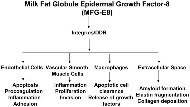
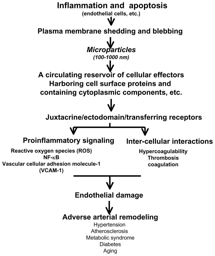
Arterial inflammation and remodeling, important sequellae of advancing age, are linked to the pathogenesis of age-associated arterial diseases e.g. hypertension, atherosclerosis, and metabolic disorders. Recently, high-throughput proteomic screening has identified milk fat globule epidermal growth factor VIII (MFG-E8) as a novel local biomarker for aging arterial walls. Additional studies have shown that MFG-E8 is also an element of the arterial inflammatory signaling network. The transcription, translation, and signaling levels of MFG-E8 are increased in aged, atherosclerotic, hypertensive, and diabetic arterial walls in vivo as well as activated vascular smooth muscle cells (VSMC) and a subset of macrophages in vitro. In VSMC, MFG-E8 increases proliferation and invasion as well as the secretion of inflammatory molecules. In endothelial cells (EC), MFG-E8 facilitates apoptosis. In addition, MFG-E8 has been found to be an essential component of the endothelial-derived microparticles that relay biosignals and modulate arterial wall phenotypes. This review mainly focuses upon the landscape of MFG-E8 expression and signaling in adverse arterial remodeling. Recent discoveries have suggested that MFG-E8 associated interventions are novel approaches for the retardation of the enhanced rates of VSMC proliferation and EC apoptosis that accompany arterial wall inflammation and remodeling during aging and age-associated arterial disease.
Conflict of interest statement
None.
Figures
References
-
- Scuteri A, Najjar SS, Muller DC, Andres R, Hougaku H, Metter EJ, Lakatta EG. Metabolic syndrome amplifies the age-associated increases in vascular thickness and stiffness. J Am Coll Cardiol. 2004 Apr 21;43(8):1388–95. - PubMed
-
- Yoshida H, Kawane K, Koike M, Mori Y, Uchiyama Y, Nagata S. Phosphatidylserine-dependent engulfment by macrophages of nuclei from erythroid precursor cells. Nature. 2005;437:754–758. - PubMed
Publication types
MeSH terms
Substances
Grants and funding
LinkOut - more resources
Full Text Sources
Other Literature Sources
Miscellaneous
