. 2012 May;20(5):1002-13.
doi: 10.1038/mt.2011.298. Epub 2012 Jan 24.

Cell fate control gene therapy based on engineered variants of human deoxycytidine kinase

Affiliations

Cell fate control gene therapy based on engineered variants of human deoxycytidine kinase

Anton Neschadim et al. Mol Ther. 2012 May.

Abstract

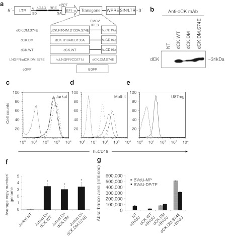
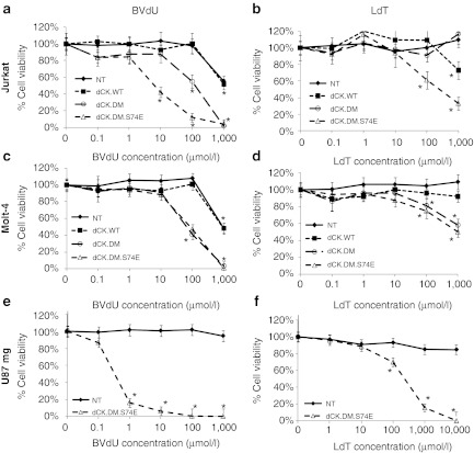
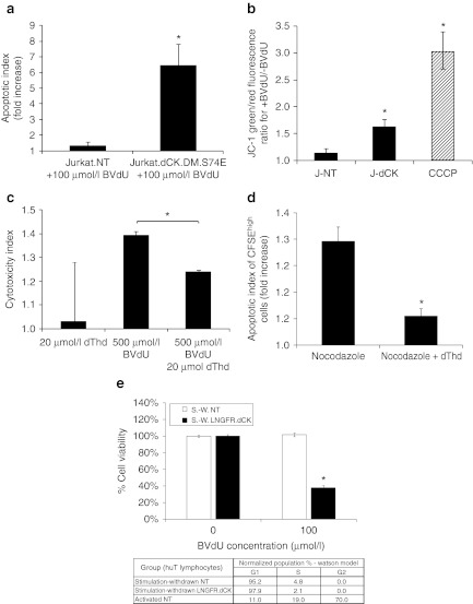
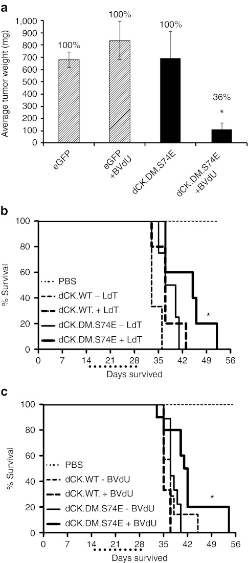
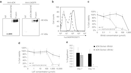
The safety of cell therapy applications can be enhanced by the introduction of Cell Fate Control (CFC) elements, which encode pharmacologically controlled cellular suicide switches. CFC Gene Therapy (CFCGT) offers the possibility of establishing control over gene-modified cells (GMCs) with regards to their proliferation, differentiation, or function. However, enzymes commonly employed in these approaches often possess poor kinetics and high immunogenicity. We describe a novel CFCGT system based on engineered variants of human deoxyCytidine Kinase (dCK) that overcomes limitations of current modalities. Mutants of dCK with rationally designed active sites that make them thymidine-activating were stably introduced into cells by recombinant lentiviral vectors (LVs). Transduced cells maintained growth kinetics and function. These dCK mutants efficiently activate bromovinyl-deoxyuridine (BVdU), L-deoxythymidine (LdT), and L-deoxyuridine (LdU), which are otherwise not toxic to wild-type cells. We show that mutant dCK-expressing Jurkat, Molt-4, and U87mg cells could be efficiently eliminated in vitro and in xenogeneic leukemia and tumor models in vivo. We also describe a fusion construct of the thymidine-activating dCK to the cytoplasmic tail-truncated LNGFR molecule and applications to in vivo eradication of primary human T cells. This novel CFCGT system offers unique plasticity with respect to the wide range of prodrugs it can potentiate, and can be used as a reliable safety switch in cell and gene therapy.


Figures

Figure 1
Figure 1
Characterization of lentiviral vector (LV)-driven dCK variant expression and prodrug activation in vitro. (a) Diagrammatic representation of LV expression constructs used in these studies. The transcription of the expression cassettes is driven off the elongation factor 1-α (EF1-α) promoter. The deoxyCytidine Kinase (dCK)-encoding vectors carry a bicistronic cassette, with the translation of the second cDNA, cytoplasmic tail-truncated human CD19 (huCD19Δ) marker, driven by the internal ribosomal entry site (IRES) derived from the encephalomyocarditis virus (EMCV). ψ, human immunodeficiency virus packaging signal; cPPT, central polypurine tract; EGFP, enhanced green fluorescent protein; LNGFRΔ, cytoplasmic tail-truncated low-affinity nerve growth factor receptor; LTR, long-terminal repeat; RRE, rev response element; SA, splice acceptor; SD, splice donor; SIN, self-inactivating LTR; WPRE, woodchuck hepatitis virus post-transcriptional regulatory element. (b) Detection of overexpressed dCK enzyme by Western blotting of lysates from transduced Jurkat cells. Next, (c) Jurkat, (d) Molt-4, and (e) U87mg cells transduced with different LV/dCK constructs and sorted by fluorescence-activated cell sorting (FACS) were analyzed by flow cytometry for human CD19 expression (dotted line, nontransduced control cells; alternating dash/dot line, dCK.WT; dashed line, dCK.DM; solid line, dCK.DM.S74E). (f) Average provirus copy numbers in sorted populations of transduced Jurkat cells were quantified by WPRE-based quantitative-PCR (Q-PCR) and standardized against a cell line known to express a single proviral copy (error bars represent SE of the mean; n = 3; *indicates statistically significant difference, P < 0.005). (g) Lysates of transduced and nontransduced control Jurkat cells treated with 10 µmol/l bromovinyl-deoxyuridine (BVdU) for 12 hours were analyzed by high-performance liquid chromatography (HPLC) for intracellular accumulation of phosphorylated BVdU metabolites, immediate dCK product BVdU-monophosphate (BVdU-MP), BVdU-diphosphate and- triphosphate (BVdU-DP/TP) (error bars represent SD; n = 3; *indicates statistically significant difference, P < 0.005).
Figure 2
Figure 2
Sensitivity of deoxyCytidine Kinase (dCK)-transduced cells to prodrug treatment in vitro. Transduced and fluorescence-activated cell sorting (FACS)-enriched (a,b) Jurkat, (c,d) Molt-4, and (e,f) U87mg cells were treated with increasing concentrations of two thymidine-based nucleoside analogues, 0.1 µmol/l–1 mmol/l bromovinyl-deoxyuridine (BVdU) (a,c,e) and 0.1 µmol/l-10 mmol/l L-deoxythymidine (LdT) (b,d,f), in culture over a period of 5 days. After 5 days, viability was assessed by the MTS assay. Corresponding mean viability is plotted for each experiment. Cells were either nontransduced (closed diamonds), or transduced with either dCK.WT (closed squares), dCK.DM (open circles), or dCK.DM.S74E (open triangles). Data shown for each group were normalized to corresponding nontreated controls (error bars represent SE of the mean; each analysis was performed at least in triplicate; *indicates statistically significant difference compared to nontreated control, P < 0.01).
Figure 3
Figure 3
Mechanisms of cell killing by activated bromovinyl-deoxyuridine (BVdU) in Jurkat cells expressing thymidine-activating deoxyCytidine Kinase (dCK). (a) Jurkat cells-expressing dCK.DM.S74E demonstrate a significant increase in the induction of apoptosis as measured by Annexin-V staining following 5 day treatment with 100 µmol/l BVdU (apoptotic index measures the fold increase in Annexin-V positive cells in BVdU-treated cells over untreated control). (b) Loss of mitochondrial inner membrane potential in Jurkat cells transduced with LV-dCK.DM.S74E and cultured in the presence or absence of 100 µmol/l BVdU for 48 hours (J-NT, nontransduced; J-dCK, dCK.DM.S74E-transduced; CCCP, 50 µmol/l carbonyl cyanide 3-chlorophenylhydrazone mitochondrial membrane potential disrupter). Increase in the ratio of green to red fluorescence of the JC-1 dye correlates with mitochondria disruption (error bars represent SD; data acquired in duplicate; *indicates statistically significant difference compared to nontransduced controls, P < 0.05). (c) Molt-4 cells transduced with dCK.DM.S74E and nontransduced controls were cultured in the presence or absence of 20 µmol/l dThd, 500 µmol/l BVdU, or both, for a period of 2 days. Viability was assessed by the MTS assay. Percent cell killing was calculated and normalized to nontreated controls. Cytotoxic index was calculated as the ratio of percent cell killing in dCK.DM.S74E-transduced cells to percent cell killing in nontransduced control cells for each treatment group (error bars represent SE of the mean; each analysis was performed in triplicate; *indicates statistically significant difference, P < 0.005). (d) Molt-4 cells transduced with dCK.DM.S74E and nontransduced controls were labeled with 10 µmol/l CFSE and cultured in the presence or absence of 100 µmol/l BVdU, and in the presence or absence of 0.1 µg/ml nocodazole, 20 µmol/l dThd, or both for 4 days. Induction of apoptosis in CFSEhigh nondividing cells was assessed by Annexin V staining. (Apoptotic index measures the fold increase in Annexin-V positive cells in BVdU-treated cells over untreated control. Error bars represent SD; data was acquired in duplicate; *indicates statistically significant differences between groups, P < 0.05). (e) Human T cells transduced with LNGFRΔ.dCK.DM.S74E vector and nontransduced controls were cultured with anti-CD3/CD28 beads and IL-2 stimulation removed (stimulation-withdrawn (S.-W.) cells). Cell-cycle distribution was assessed by propidium iodide staining of fixed cells, and analyzed by flow cytometry, with the results tabulated. Cell viability following a 4-day exposure of the cells to 100 µmol/l BVdU was assessed by the MTS assay. (Error bars represent SE; data acquired in quadruplicate; *indicates statistically significant differences between BVdU-treated and untreated groups, P < 0.01).
Figure 4
Figure 4
U87mg and Molt-4 cells transduced with the thymidine-activating mutant of deoxyCytidine Kinase (dCK) are sensitive to prodrug-mediated cell killing in vivo in tumor and leukemia models of cancer. (a) Subcutaneous tumors in NOD/SCID mice formed by U87mg cells transduced with dCK.DM.S74E but not control eGFP-transduced cells were efficiently eradicated in vivo by treatment with bromovinyl-deoxyuridine (BVdU) (eGFP, U87mg cells transduced with LV-eGFP; dCK.DM.S74E, U87mg cells transduced with LV-dCK.DM.S74E; +BVdU, treated with 60 mg/kg/day of BVdU for 21 days). (Error bars represent SE of the mean; percentages above the bars represent proportion of mice that had measurable tumors; n = 7–11 for treatment groups and n = 3 for PBS controls; *indicates statistically significant difference, P <0.005). (b,c) Kaplan-Meier survival curves of mice with Molt-4-derived leukemias. Aggressive T cell leukemia was induced in pre-conditioned NOD/SCID animals by intravenous tail vein administration of human Molt-4 cells (dCK.WT, Molt-4 cells transduced with LV-dCK.WT; dCK.DM.S74E, Molt-4 cell transduced with LV-dCK.DM.S74E; PBS, PBS-injected control animals; +LdT, treated with 50 mg/kg/day of L-deoxythymidine (LdT); +BVdU, treated with 50 mg/kg/day of BVdU; dotted line below x-axis underlines the drug treatment window). Survival of mice with established leukemias derived from Molt-4 cells-expressing dCK.DM.S74E was increased with LdT (b) or BVdU (c) treatments (median survival increase of 1 week) administered for 14 days starting day 15 after cell injection (n = 3–10; *indicates statistically significant difference compared to control, P < 0.05). LV, lentiviral vector.
Figure 5
Figure 5
Primary human T lymphocytes transduced with the LNGFR-deoxyCytidine Kinase (dCK) fusion are sensitive to prodrug-mediated cell killing in vitro. (a) Cell lysates from Jurkat cells transduced with LVs engineering the expression of either the dCK.DM.S74E or the LNGFRΔ-dCK.DM.S74E were probed by western blotting for the expression of dCK (left panel) and LNGFR (right panel). (b) Nontransduced (dotted line) and LNGFRΔ-dCK.DM.S74E-transduced human T cells were analyzed by flow cytometry for the expression of huLNGFR, before (dashed line) and after (solid line) magnetic enrichment. LNGFRΔ-dCK.DM.S74E-transduced (dashed line) and nontransduced (solid line) human T cells were treated or not with increasing concentrations of (c) bromovinyl-deoxyuridine (BVdU) or (d) L-deoxythymidine (LdT) in culture over a period of 5 days. Viability was then assessed by the MTS assay. Corresponding mean viability is plotted for each experiment (error bars represent SE of the mean; each analysis was performed in triplicate; *indicates significant difference compared to control, P < 0.01). (d) 2 × 10⁶ LNGFRΔ-dCK.DM.S74E-transduced human T cells were infused into pre-conditioned (day 0) NOD/SCID animals by intravenous tail vein administration (day 1). Animals were treated (shaded bars) with 50 mg/kg/day of BVdU on days 2, 3, 5, and 6, and then with 80 mg/kg/day for days 8–14, or left untreated (black bars). Percent engraftment of human T cells was measured by simultaneous staining of peripheral blood cells with antihuman CD45 and anti-LNGFR antibodies. (n = 4; *indicates statistically significant difference compared to untreated controls, P < 0.05).

Similar articles

Cited by

References

    1. Rezvani AR., and, Storb RF. Separation of graft-vs.-tumor effects from graft-vs.-host disease in allogeneic hematopoietic cell transplantation. J Autoimmun. 2008;30:172–179. - PMC - PubMed
    1. Fraser CJ, Hirsch BA, Dayton V, Creer MH, Neglia JP, Wagner JE.et al. (2005First report of donor cell-derived acute leukemia as a complication of umbilical cord blood transplantation Blood 1064377–4380. - PMC - PubMed
    1. Amariglio N, Hirshberg A, Scheithauer BW, Cohen Y, Loewenthal R, Trakhtenbrot L.et al. (2009Donor-derived brain tumor following neural stem cell transplantation in an ataxia telangiectasia patient PLoS Med 6e1000029. - PMC - PubMed
    1. Cooley LD, Sears DA, Udden MM, Harrison WR., and, Baker KR. Donor cell leukemia: report of a case occurring 11 years after allogeneic bone marrow transplantation and review of the literature. Am J Hematol. 2000;63:46–53. - PubMed
    1. Goring DR., and, DuBow MS. A cytotoxic effect associated with 9-(1,3-dihydroxy-2-propoxymethyl)-guanine is observed during the selection for drug resistant human cells containing a single herpesvirus thymidine kinase gene. Biochem Biophys Res Commun. 1985;133:195–201. - PubMed

Publication types

MeSH terms