

Soluble vascular endothelial growth factor decoy receptor FP3 exerts potent antiangiogenic effects
- PMID: 22273580
- PMCID: PMC3345977
- DOI: 10.1038/mt.2011.285
Soluble vascular endothelial growth factor decoy receptor FP3 exerts potent antiangiogenic effects
Abstract
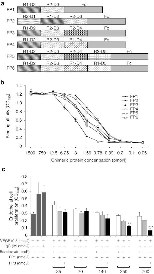
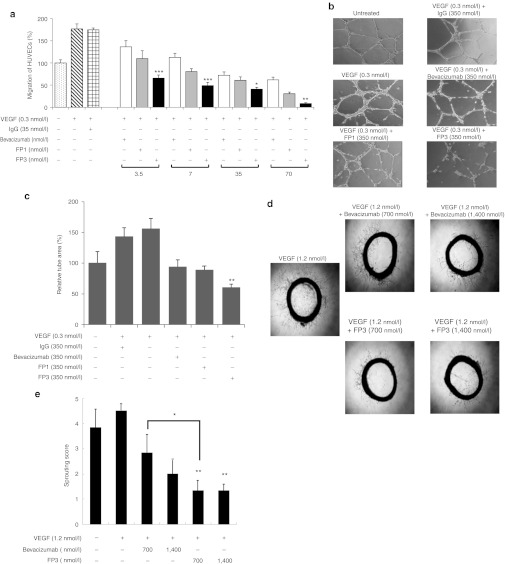
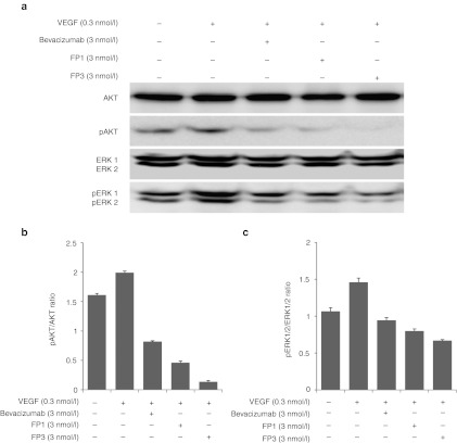
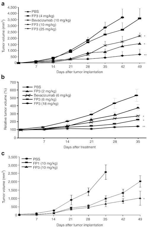
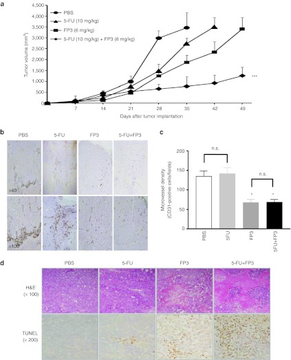
The binding of vascular endothelial growth factor (VEGF) to its receptors stimulates tumor growth; therefore, modulation of VEGF would be a viable approach for antiangiogenic therapy. We constructed a series of soluble decoy receptors containing different VEGF receptor 1 (FLT1) and VEGF receptor 2 (KDR) extracellular domains fused with the Fc region of human immunoglobulin (Ig) and evaluated their antiangiogenic effects and antitumor effects. Results of in vitro binding and cell proliferation assays revealed that decoy receptor FP3 had the highest affinity to VEGF-A and -B. Compared with bevacizumab, FP3 more effectively inhibited human umbilical vein endothelial cell (HUVEC) migration and vessel sprouting from rat aortic rings. FP3 significantly reduced phosphorylation of AKT and ERK1/2, critical proteins in the VEGF-mediated survival pathway in endothelial cells. Moreover, FP3 inhibited tumor growth in human hepatocellular carcinoma (HepG2), breast cancer (MCF-7), and colorectal cancer (LoVo) tumor models, and reduced microvessel density in tumor tissues. The FP3-mediated inhibition of tumor growth was significantly higher than that of bevacizumab at the same dose. FP3 also demonstrated synergistic antitumor effects when combined with 5-fluorouracil (5-FU). Taken together, FP3 shows a high affinity for VEGF and produced antiangiogenic effects, suggesting its potential for treating angiogenesis-related diseases such as cancer.
Figures
Similar articles
-
FP3: a novel VEGF blocker with antiangiogenic effects in vitro and antitumour effects in vivo.Clin Transl Oncol. 2011 Dec;13(12):878-84. doi: 10.1007/s12094-011-0749-z. Clin Transl Oncol. 2011. PMID: 22126731
-
CEP-7055: a novel, orally active pan inhibitor of vascular endothelial growth factor receptor tyrosine kinases with potent antiangiogenic activity and antitumor efficacy in preclinical models.Cancer Res. 2003 Sep 15;63(18):5978-91. Cancer Res. 2003. PMID: 14522925
-
Potent and long-term antiangiogenic efficacy mediated by FP3-expressing oncolytic adenovirus.Int J Cancer. 2015 Nov 1;137(9):2253-69. doi: 10.1002/ijc.29592. Epub 2015 May 26. Int J Cancer. 2015. PMID: 25944623
-
Antitumor effects of FP3 in combination with capecitabine on PDTT xenograft models of primary colon carcinoma and related lymphatic and hepatic metastases.Cancer Biol Ther. 2012 Jul;13(9):737-44. doi: 10.4161/cbt.20556. Epub 2012 May 23. Cancer Biol Ther. 2012. PMID: 22617773
-
Use of soluble recombinant decoy receptor vascular endothelial growth factor trap (VEGF Trap) to inhibit vascular endothelial growth factor activity.Clin Colorectal Cancer. 2004 Oct;4 Suppl 2:S81-5. doi: 10.3816/ccc.2004.s.013. Clin Colorectal Cancer. 2004. PMID: 15479484 Review.
Cited by
-
Targeting tumor vasculature through oncolytic virotherapy: recent advances.Oncolytic Virother. 2015 Nov 11;4:169-81. doi: 10.2147/OV.S66045. eCollection 2015. Oncolytic Virother. 2015. PMID: 27512680 Free PMC article. Review.
-
Anti-angiogenic effect of KH902 on retinal neovascularization.Graefes Arch Clin Exp Ophthalmol. 2013 Sep;251(9):2131-9. doi: 10.1007/s00417-013-2392-6. Epub 2013 Jun 6. Graefes Arch Clin Exp Ophthalmol. 2013. PMID: 23740520
-
SARI inhibits angiogenesis and tumour growth of human colon cancer through directly targeting ceruloplasmin.Nat Commun. 2016 Jun 29;7:11996. doi: 10.1038/ncomms11996. Nat Commun. 2016. Retraction in: Nat Commun. 2023 Jul 20;14(1):4377. doi: 10.1038/s41467-023-40067-6. PMID: 27353863 Free PMC article. Retracted.
-
Profile of conbercept in the treatment of neovascular age-related macular degeneration.Drug Des Devel Ther. 2015 Apr 22;9:2311-20. doi: 10.2147/DDDT.S67536. eCollection 2015. Drug Des Devel Ther. 2015. PMID: 25960634 Free PMC article. Review.
-
Intracellular virion traffic to the endosome driven by cell type specific sialic acid receptors determines parvovirus tropism.Front Microbiol. 2023 Jan 23;13:1063706. doi: 10.3389/fmicb.2022.1063706. eCollection 2022. Front Microbiol. 2023. PMID: 36756201 Free PMC article.
References
-
- Hanahan D., and, Folkman J. Patterns and emerging mechanisms of the angiogenic switch during tumorigenesis. Cell. 1996;86:353–364. - PubMed
-
- Ferrara N., and, Alitalo K. Clinical applications of angiogenic growth factors and their inhibitors. Nat Med. 1999;5:1359–1364. - PubMed
-
- Yancopoulos GD, Davis S, Gale NW, Rudge JS, Wiegand SJ., and, Holash J. Vascular-specific growth factors and blood vessel formation. Nature. 2000;407:242–248. - PubMed
Publication types
MeSH terms
Substances
LinkOut - more resources
Full Text Sources
Other Literature Sources
Miscellaneous
