. 2012 Jan 24:3:639.
doi: 10.1038/ncomms1647.

Three-dimensional analysis of ribonucleoprotein complexes in influenza A virus

Affiliations
Free PMC article

Three-dimensional analysis of ribonucleoprotein complexes in influenza A virus

Takeshi Noda et al. Nat Commun. .
Free PMC article

Abstract

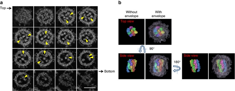
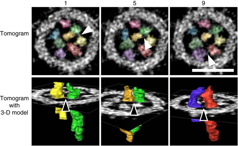
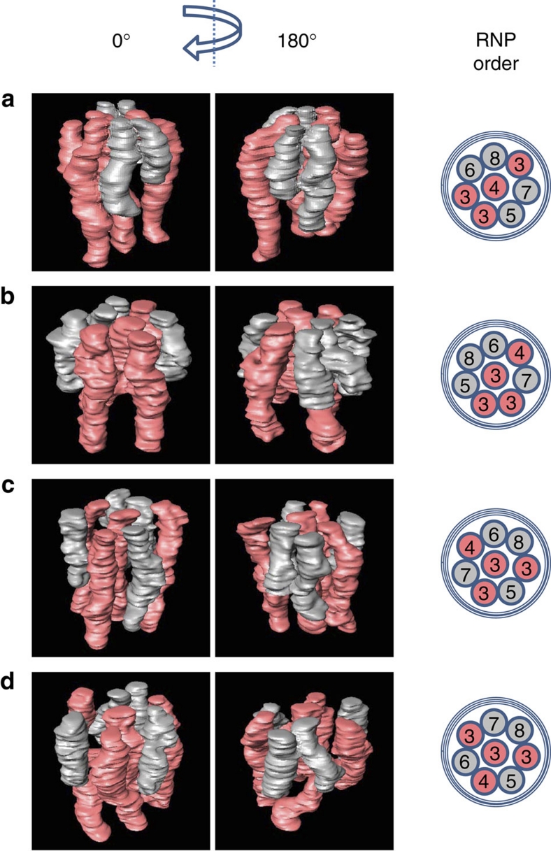
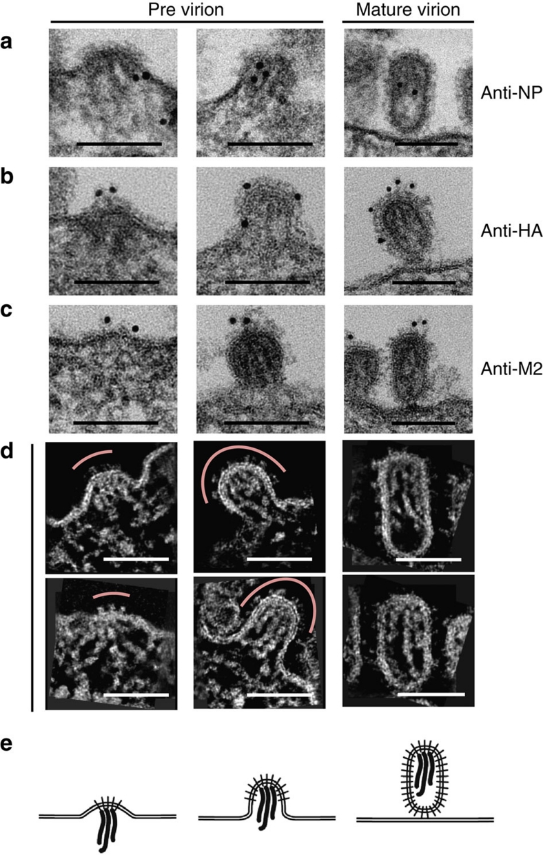
The influenza A virus genome consists of eight single-stranded negative-sense RNA (vRNA) segments. Although genome segmentation provides advantages such as genetic reassortment, which contributes to the emergence of novel strains with pandemic potential, it complicates the genome packaging of progeny virions. Here we elucidate, using electron tomography, the three-dimensional structure of ribonucleoprotein complexes (RNPs) within progeny virions. Each virion is packed with eight well-organized RNPs that possess rod-like structures of different lengths. Multiple interactions are found among the RNPs. The position of the eight RNPs is not consistent among virions, but a pattern suggests the existence of a specific mechanism for assembly of these RNPs. Analyses of budding progeny virions suggest two independent roles for the viral spike proteins: RNP association on the plasma membrane and the subsequent formation of the virion shell. Our data provide further insights into the mechanisms responsible for segmented-genome packaging into virions.


Figures

Figure 1
Figure 1. Dual-axis STEM tomography of a progeny influenza A virion.
(a) Consecutive transverse sections of a progeny virion (0.5 nm thick at 5.0-nm intervals) from its top to bottom. The RNPs, approximately 12 nm in diameter, are present within the viral envelope. Representative associations between the RNPs are shown as examples (arrowheads). Scale bar, 50 nm. (b) A 3-D model of the eight RNPs within a virion. Each RNP and the viral envelope are represented by a different colour.
Figure 2
Figure 2. Linkages among the eight RNPs in a virion.
Short string-like structures of approximately 2 nm in diameter can be seen between the RNPs (arrowheads). Tomograms, 0.5-nm thick, are shown in the upper row. The 3-D models of two RNPs connected by a string-like structure are shown in the lower row. Virtual slice numbers (1, 5 and 9) of 0.5-nm thick tomograms are given above the tomograms. The colour scheme of the RNPs is the same as that shown in Figure 1b. Scale bar, 50 nm.
Figure 3
Figure 3. 3-D models of the eight RNPs in different virions.
(a?d) Four examples of 3-D models of the multi-segment complex are shown. The schemes illustrated in the right column show the order of the eight RNPs, which are numbered from 3 to 8 in descending order of length (that is, 3, 3, 3, 4, 5, 6, 7 and 8). For clarity, the four longer RNPs (that is, numbers 3, 3, 3 and 4) are coloured red and the four shorter RNPs (that is, numbers 5, 6, 7 and 8) are coloured grey.
Figure 4
Figure 4. Packaging of RNPs into the virion.
The left column shows the initial stage of budding, the middle column shows the middle stage and the right column shows mature virions. (a) Immunostaining with an anti-NP antibody conjugated with 10 nm gold beads. (b) Immunostaining with an anti-HA antibody conjugated with 10 nm gold beads. (c) Immunostaining with an anti-M2 antibody conjugated with 10 nm gold beads. (d) Slab slices (10 nm) of previrions in the process of budding and mature virions. Red lines in the left and middle columns indicate the membrane region where viral spike proteins are present. Scale bar, 100 nm. (e) Schematic diagrams of the RNP packaging process. Initial (left), middle (center) and late (right) steps of budding.

Similar articles

Cited by

References

    1. Palese P in Fields Virology (eds Knipe, D. M. & Howley, P. M.) 1647–1689 (Lippincott, Williams & Wilkins, Philadelphia, 2007).
    1. Compans R. W., Content J. & Duesberg P. H. Structure of ribonucleoprotein of influenza virus. J. Virol. 10, 795–800 (1972). - PMC - PubMed
    1. Jennings P. A., Finch J. T., Winter G. & Robertson J. S. Does the higher order structure of the influenza virus ribonucleoprotein guide sequence rearrangements in influenza viral RNA? Cell 34, 619–627 (1983). - PubMed
    1. Pons M. W., Schulze I. T., Hirst G. K. & Hauser R. Isolation and characterization of the ribonucleoprotein oh influenza virus. Virology. 39, 250–259 (1969). - PubMed
    1. Ortega J. et al.. Ultrastructural and functional analyses of recombinant influenza virus ribonucleoproteins suggest dimerization of nucleoprotein during virus amplification. J. Virol. 74, 156–163 (2000). - PMC - PubMed

Publication types