. 2012 Apr;126(2):554-68.
doi: 10.1093/toxsci/kfs024. Epub 2012 Jan 23.

Aryl hydrocarbon receptor (AhR) regulates silica-induced inflammation but not fibrosis

Affiliations

Aryl hydrocarbon receptor (AhR) regulates silica-induced inflammation but not fibrosis

Celine A Beamer et al. Toxicol Sci. 2012 Apr.

Abstract

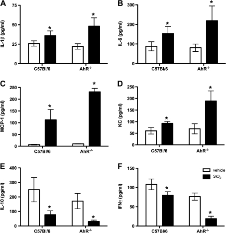
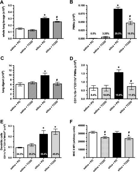
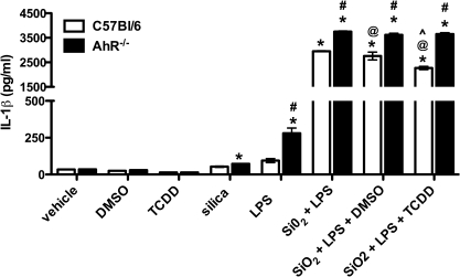
The aryl hydrocarbon receptor (AhR), a ligand-activated transcription factor, is responsible for mediating a variety of pharmacological and toxicological effects caused by halogenated aromatic hydrocarbons such as 2,3,7,8-tetrachlorodibenzo-p-dioxin (TCDD). However, recent evidence has revealed that the AhR also has numerous physiological roles aside from xenobiotic metabolism, including regulation of immune and inflammatory signaling as well as normal development and homeostasis of several organs. To investigate the role of the AhR in crystalline silica (SiO(2))-induced inflammation and fibrosis, C57Bl/6 and AhR(-/)(-) mice were exposed to SiO(2) or vehicle. Similarly, C57Bl/6 mice were exposed to SiO(2) and TCDD either simultaneously or sequentially to assess whether AhR activation alters inflammation and fibrosis. SiO(2)-induced acute lung inflammation was more severe in AhR(-)(/-) mice; however, the fibrotic response of AhR(-)(/-) mice was attenuated compared with C57Bl/6 mice. In a model of chronic SiO(2) exposure, AhR activation by TCDD in C57Bl/6 mice resulted in reduced inflammation; however, the fibrotic response was not affected. Bone marrow-derived macrophages (BMM) from AhR(-)(/-) mice also produced higher levels of cytokines and chemokines in response to SiO(2). Analysis of gene expression revealed that BMM derived from AhR(-)(/-) mice exhibit increased levels of pro-interleukin (IL)-1β, IL-6, and Bcl-2, yet decreased levels of signal transducers and activators of transcription (STAT)2, STAT5a, and serpin B2 (Pai-2) in response to SiO(2).


Figures

FIG. 1.
FIG. 1.
Cellular response to SiO2 exposure in AhR/− and C57Bl/6 mice. Crystalline silica (SiO2) increased the number of cells recovered via WLL in AhR/− compared with C57Bl/6 mice (A). Alveolar inflammation dominated by PMNs was observed—a response that was augmented in SiO2-exposed AhR/− mice (B); n = 5–7, values are means ± SEM; *p < 0.05 compared with wild-type mice.
FIG. 2.
FIG. 2.
Expression of cytokines and chemokines in SiO2-exposed AhR/− and C57Bl/6 mice 3 days postinstillation. Levels of IL-1β, IL-6, MCP-1, and KC were increased, whereas levels of IL-10 and IFNγ were decreased in the WLL fluid after SiO2 exposure in AhR/− and wild-type C57Bl/6 mice. These responses were exacerbated in AhR/− mice compared with wild-type mice; n = 5–8, values are means ± SEM; *p < 0.05 compared with vehicle control.
FIG. 3.
FIG. 3.
Innate immune responses to SiO2 exposure in TCDD-induced AhR–activated C57Bl/6 mice. Chronic SiO2 exposure increased the total number of lavageable cells (A), and interstitial leukocytes recovered from the lung (C). This increased cellularity was reduced by concomitant activation of the AhR. Chronic SiO2 exposure increased the percent and absolute number of neutrophils (PMNs) in the WLL (B) and LD (D)—events that were inhibited by TCDD-induced AhR activation. Although the percent and absolute number of interstitial DCs were increased in response to chronic SiO2 exposure, neither parameter was affected by AhR activation (E). However, TCDD attenuated MHC class II expression on interstitial DCs (F); n = 5–6, values are means ± SEM; *p < 0.05 compared with vehicle.
FIG. 4.
FIG. 4.
TCDD-induced AhR activation attenuated inflammation but not fibrosis in C57Bl/6 mice. Inflammation and fibrosis were evaluated by Gomori's trichrome staining of lung sections. Representative sections from vehicle control mice, e.g., saline + peanut oil (A) and saline + TCDD (C) show normal tissue and cell architecture and no inflammatory or fibrotic response (arrows). In contrast, chronic SiO2 exposure results in enhanced infiltration of inflammatory cells and collagen deposition (arrows) (B). In comparison, TCDD-induced AhR activation decreased the inflammatory infiltrate observed but had no effect on fibrosis (arrows) (D).
FIG. 5.
FIG. 5.
Inflammatory cytokine response to SiO2 exposure following AhR activation. Levels of IL-1β (A), IL-6 (B), and IL-22 (C) were evaluated in the WLL fluid 12 weeks after the initial SiO2 exposure in C57Bl/6 mice. Both sequential and concomitant TCDD-induced AhR activation reduced production of IL-1β and increased IL-6 and IL-22. TCDD alone had no effect on IL-1β or IL-6 secretion but increased IL-22 levels; n = 5–6, values are means ± SEM; *p < 0.05 compared with vehicle control, #p < 0.05 compared with silica + peanut oil.
FIG. 6.
FIG. 6.
Cytokine response in BMM derived from wild-type and AhR/− mice. Twenty-four hours following SiO2 + LPS treatment, the levels of IL-1β (A), IL-6 (B), MCP-1 (C), KC (D), and IFNγ were significantly increased in tissue culture supernatants. AhR/− BMM showed enhanced levels of inflammatory cytokines and chemokines compared with wild-type mice. These responses were inhibited by 4 h pretreatment with 20 ng/ml recombinant mouse IL-10; n = 3–5, values are means ± SEM; *p < 0.05 compared with vehicle control. #p < 0.05 compared to SiO2 + LPS.
FIG. 7.
FIG. 7.
IL-1β production is attenuated in BMM following AhR activation with TCDD. Activation of the inflammasome following stimulation of BMM with SiO2 plus LPS for 24 h resulted in elevated levels of IL-1β present in tissue culture supernatants. This response was reduced by concomitant treatment with 10nM TCDD in wild-type macrophages. In contrast, no change was observed in the levels of IL-1β present in tissue culture supernatants in BMM derived from AhR/− mice; n = 3, values are means ± SEM; *p < 0.05 compared with vehicle control, #p < 0.05 AhR/− compared with wild type, @p < 0.05 compared with silica + LPS, ^p < 0.05 compared with SiO2 + LPS + DMSO.

References

    1. Bankoti J, Burnett A, Navarro S, Miller AK, Rase B, Shepherd DM. Effects of TCDD on the fate of naive dendritic cells. Toxicol. Sci. 2010a;115:422–434. - PMC - PubMed
    1. Bankoti J, Rase B, Simones T, Shepherd DM. Functional and phenotypic effects of AhR activation in inflammatory dendritic cells. Toxicol. Appl. Pharmacol. 2010b;246:18–28. - PMC - PubMed
    1. Barbarin V, Nihoul A, Misson P, Arras M, Delos M, Leclercq I, Lison D, Huaux F. The role of pro- and anti-inflammatory responses in silica-induced lung fibrosis. Respir. Res. 2005;6:112. - PMC - PubMed
    1. Beamer CA, Holian A. Scavenger receptor class A type I/II (CD204) null mice fail to develop fibrosis following silica exposure. Am. J. Physiol. Lung Cell. Mol. Physiol. 2005;289:L186–L195. - PubMed
    1. Beamer CA, Holian A. Antigen presenting cell population dynamics during murine silicosis. Am. J. Respir. Cell Mol. Biol. 2007;37:729–738. - PMC - PubMed

Publication types

MeSH terms

Substances