

Changes of ribosomal protein S3 immunoreactivity and its new expression in microglia in the mice hippocampus after lipopolysaccharide treatment
- PMID: 22274408
- PMCID: PMC11498381
- DOI: 10.1007/s10571-012-9802-x
Changes of ribosomal protein S3 immunoreactivity and its new expression in microglia in the mice hippocampus after lipopolysaccharide treatment
Abstract
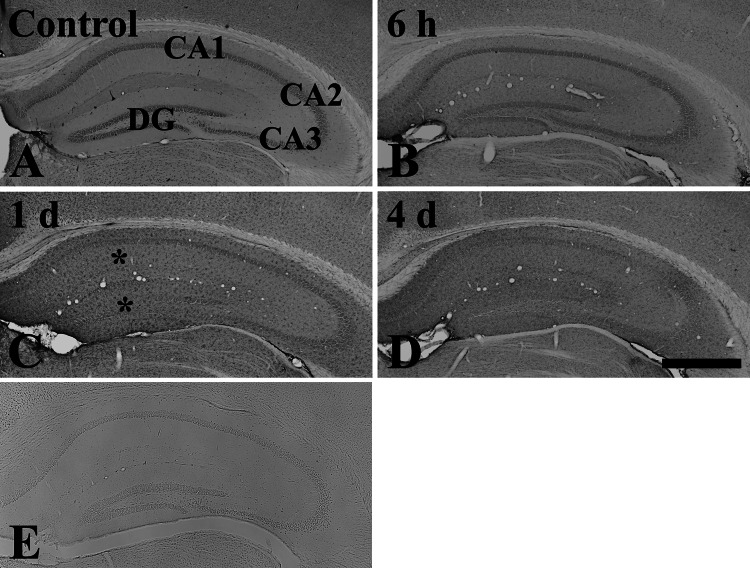
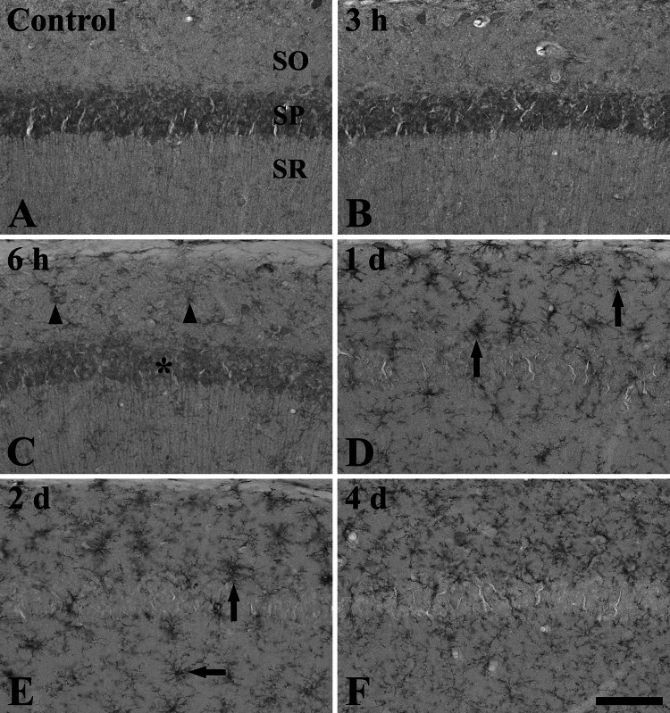
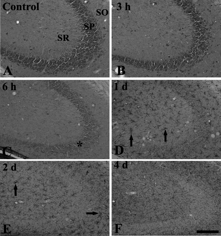
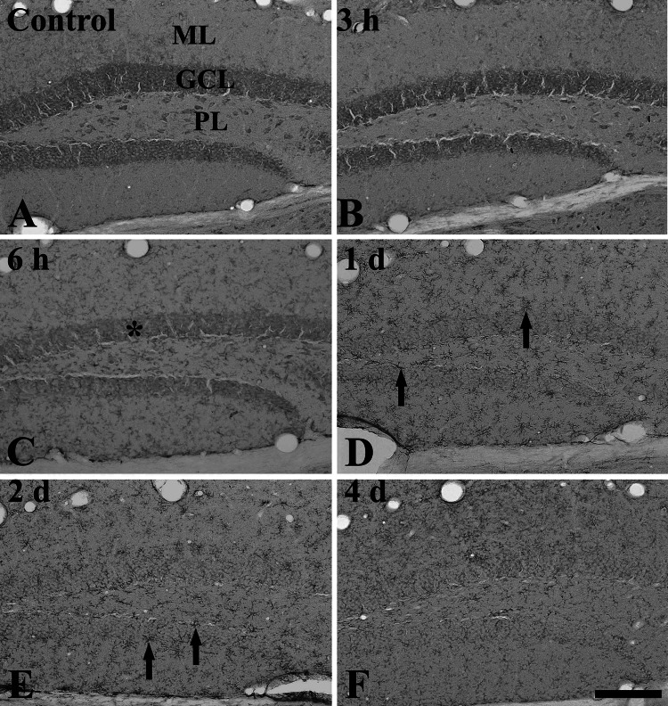
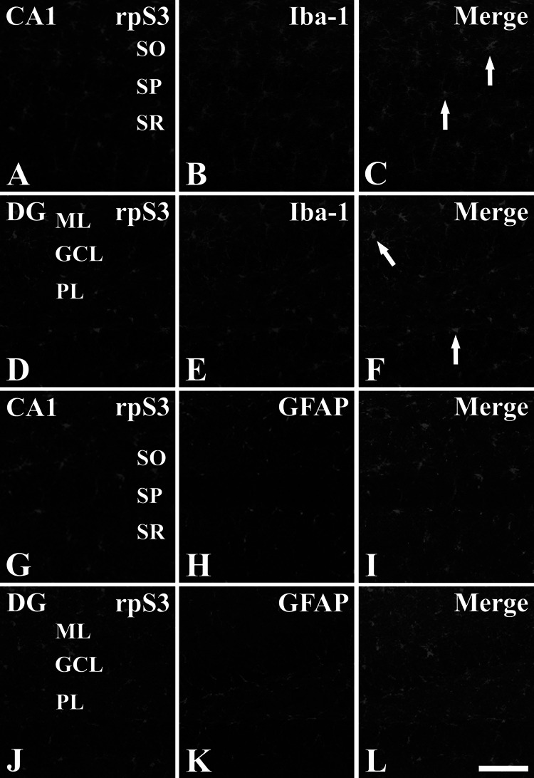
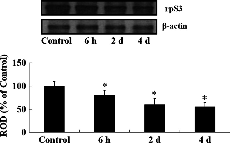
Lipopolysaccharide (LPS) has been used as a reagent for a model of systemic inflammatory response. Ribosomal protein S3 (rpS3) is a multi-functional protein that is involved in transcription, metastasis, DNA repair, and apoptosis. In the present study, we examined the changes of rpS3 immunoreactivity in the mouse hippocampus after systemic administration of 1 mg/kg of LPS. From 6 h after LPS treatment, rpS3 immunoreactivity was decreased in pyramidale cells of the hippocampus proper (CA1-CA3 regions) and in granule cells of the dentate gyrus. At this point in time, rpS3 immunoreactivity began to increase in non-pyramidal cells and non-granule cells. From 1 day after LPS treatment, rpS3 immunoreactivity in pyramidal and granule cells was hardly detected; however, strong rpS3 immunoreactivity was shown in non-pyramidal and non-granule cells. Based on double immunofluorescence staining for rpS3/ionized calcium-binding adapter 1 (Iba-1, a marker for microglia) and glial fibrillary acidic protein (GFAP, a marker for astrocytes), strong rpS3 immunoreactivity was expressed in Iba-1-immunoreactive microglia, not in GFAP-immunoreactive astrocytes, at 1 and 2 days after LPS treatment. These results indicate that rpS3 immunoreactivity changes only in pyramidal and granule cells, and rpS3 is expressed only in activated microglia after LPS treatment: this may be associated with the neuroinflammatory responses in the brain.
Figures
Similar articles
-
Ribosomal protein s3 immunoreactivity in the young, adult and aged gerbil hippocampus.J Vet Med Sci. 2011 Mar;73(3):361-5. doi: 10.1292/jvms.10-0247. Epub 2010 Oct 15. J Vet Med Sci. 2011. PMID: 20962462
-
Changes in ribosomal protein S3 immunoreactivity and its protein levels in the gerbil hippocampus following subacute and chronic restraint stress.Neurochem Res. 2012 Jul;37(7):1428-35. doi: 10.1007/s11064-012-0727-z. Epub 2012 Mar 6. Neurochem Res. 2012. PMID: 22392256
-
Systemic administration of lipopolysaccharide induces cyclooxygenase-2 immunoreactivity in endothelium and increases microglia in the mouse hippocampus.Cell Mol Neurobiol. 2010 May;30(4):531-41. doi: 10.1007/s10571-009-9477-0. Epub 2009 Nov 12. Cell Mol Neurobiol. 2010. PMID: 19908141 Free PMC article.
-
Alterations in microglial phenotype and hippocampal neuronal function in transgenic mice with astrocyte-targeted production of interleukin-10.Brain Behav Immun. 2015 Mar;45:80-97. doi: 10.1016/j.bbi.2014.10.015. Epub 2014 Oct 31. Brain Behav Immun. 2015. PMID: 25449577
-
Alterations of glial cells in the mouse hippocampus during postnatal development.Cell Mol Neurobiol. 2009 Dec;29(8):1181-9. doi: 10.1007/s10571-009-9412-4. Cell Mol Neurobiol. 2009. PMID: 19472050 Free PMC article.
Cited by
-
Amitriptyline improves motor function via enhanced neurotrophin signaling and mitochondrial functions in the murine N171-82Q Huntington disease model.J Biol Chem. 2015 Jan 30;290(5):2728-43. doi: 10.1074/jbc.M114.588608. Epub 2014 Dec 11. J Biol Chem. 2015. PMID: 25505248 Free PMC article.
-
Change of calbindin D-28k protein expression in the mice hippocampus after lipopolysaccharide treatment.J Vet Med Sci. 2015 Mar;77(3):349-52. doi: 10.1292/jvms.14-0509. Epub 2014 Nov 26. J Vet Med Sci. 2015. PMID: 25428702 Free PMC article.
References
-
- Ahn EH, Kim DW, Kang HW, Shin MJ, Won MH, Kim J, Kim DJ, Kwon OS, Kang TC, Han KH, Park J, Eum WS, Choi SY (2010) Transduced PEP-1-ribosomal protein S3 (rpS3) ameliorates 12-O-tetradecanoylphorbol-13-acetate-induced inflammation in mice. Toxicology 276:192–197 - PubMed
-
- Aloisi F (2001) Immune function of microglia. Glia 36:165–179 - PubMed
-
- Ambrosini E, Aloisi F (2004) Chemokines and glial cells: a complex network in the central nervous system. Neurochem Res 29:1017–1038 - PubMed
Publication types
MeSH terms
Substances
LinkOut - more resources
Full Text Sources
Miscellaneous
