Review
. 2012 May;29(5):1180-8.
doi: 10.1007/s11095-012-0679-7. Epub 2012 Jan 25.

Magnetic nanoparticles for cancer diagnosis and therapy

Affiliations
Review

Magnetic nanoparticles for cancer diagnosis and therapy

Mehmet V Yigit et al. Pharm Res. 2012 May.

Abstract

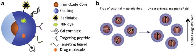
Nanotechnology is evolving as a new field that has a potentially high research and clinical impact. Medicine, in particular, could benefit from nanotechnology, due to emerging applications for noninvasive imaging and therapy. One important nanotechnological platform that has shown promise includes the so-called iron oxide nanoparticles. With specific relevance to cancer therapy, iron oxide nanoparticle-based therapy represents an important alternative to conventional chemotherapy, radiation, or surgery. Iron oxide nanoparticles are usually composed of three main components: an iron core, a polymer coating, and functional moieties. The biodegradable iron core can be designed to be superparamagnetic. This is particularly important, if the nanoparticles are to be used as a contrast agent for noninvasive magnetic resonance imaging (MRI). Surrounding the iron core is generally a polymer coating, which not only serves as a protective layer but also is a very important component for transforming nanoparticles into biomedical nanotools for in vivo applications. Finally, different moieties attached to the coating serve as targeting macromolecules, therapeutics payloads, or additional imaging tags. Despite the development of several nanoparticles for biomedical applications, we believe that iron oxide nanoparticles are still the most promising platform that can transform nanotechnology into a conventional medical discipline.


Figures

Fig. 1
Fig. 1
(a) The magnetic nanoparticle is composed of magnetic core/cores around which there is a coating. The coating is complexed with different moieties for additional functionalities. (b) Superparamagnetic iron oxide nanoparticles have a single magnetic domain because of their small magnetic core and are greatly magnetized under an externally applied magnetic field.
Fig. 2
Fig. 2
(a) Magnetic nanoparticles functionalized with siRNA targeting GFP expression (MN-NIRF-siGFP). (b) In vitro testing of MN-NIRF-siGFP cell uptake and silencing efficiency in stably transfected 9 L-GFP gliosarcoma cells. (b) The silencing effect was manifested as a concentration-dependent decrease in GFP relative fluorescence levels. Data represent an average of three experiments. RFU, relative fluorescence units. c Confocal microscopy showing MN-NIRF-siGFP accumulation in 9 L-GFP and 9 L-RFP cells. Note that although the probe accumulated in both cell lines (Cy5.5 fluorescence, blue), there was a substantial silencing effect in the 9 L-GFP cell line, resulting in marked suppression of GFP fluorescence. Scale bar, 20 μm. (d) In vivo imaging of MN-NIRF-siGFP silencing in tumors. (a) In vivo NIRF optical imaging of mice bearing bilateral 9 L-GFP and 9 L-RFP tumors 48 h after intravenous probe injection. There was a marked decrease in 9 L-GFP–associated fluorescence (P=0.0083) and no change in 9 L-RFP fluorescence. To generate GFP/RFP reconstructions, GFP and RFP images were acquired separately and then merged. (Reprinted with permission from Ref by Nature Publishing Group).
Fig. 3
Fig. 3
(a) Schematic illustration of anti HER2/neu antibody-modified pH-sensitive drug-releasing magnetic nanoparticles (HER-DMNPs) for cancer therapy followed by MRI. (b) Color-coded T 2-weighted MR images of tumor-bearing mice after the intravenous injection of HER-DMNPs and IRR-DMNPs at various time intervals, respectively. Tumor regions are indicated with a white dashed boundary. (c) Comparative therapeutic efficacy study in the in vivo model. (black circle: HER-DMNPs, dark gray triangle: IRR-DMNPs, gray square: DOX, and white diamond: saline). Black arrow indicates the day of cancer cells (NIH3T6.7 cells) implantation in mice. (Reprinted with permission from Ref by John Wiley and Sons.)
Fig. 4
Fig. 4
(a) Monocytes are obtained by cytopheresis from stage-III melanoma patients. (b) They are cultured and labeled with SPIO particles and 111In. (c,d) The cells are then injected intranodally into a (either cervical, inguinal or axillary) lymph node basin that is to be resected and their biodistribution is monitored in vivo by scintigraphy (c) and MRI at 3 Tesla (d). (eg) The lymph node basis is resected and separate lymph nodes are visualized with high resolution MRI at 7 Tesla (f) and histology (g). (Reprinted with permission from Ref by Nature Publishing Group.)

Similar articles

Cited by

References

    1. Pautler M, Brenner S. Nanomedicine: promises and challenges for the future of public health. Int J Nanomedicine. 2010;5:803–9. - PMC - PubMed
    1. Kievit FM, Zhang M. Surface engineering of iron oxide nanoparticles for targeted cancer therapy. Acc Chem Res. 2011;44:853–62. - PMC - PubMed
    1. Gupta AK, Wells S. Surface-modified superparamagnetic nanoparticles for drug delivery: preparation, characterization, and cytotoxicity studies. IEEE Trans NanoBiosci. 2004;3:66–73. - PubMed
    1. Pankhurst QA, Connolly J, Jones SK, Dobson J. Applications of magnetic nanoparticles in biomedicine. J Phys D: Appl Phys. 2003;36:R167–81.
    1. Anzai Y, Piccoli CW, Outwater EK, et al. Evaluation of neck and body metastases to nodes with ferumoxtran 10-enhanced MR imaging: phase III safety and efficacy study. Radiology. 2003;228:777–88. - PubMed