

Frontal sinus osteoma with osteoblastoma-like histology and associated intracranial pneumatocele
- PMID: 22274656
- PMCID: PMC3422592
- DOI: 10.1007/s12105-012-0332-0
Frontal sinus osteoma with osteoblastoma-like histology and associated intracranial pneumatocele
Abstract
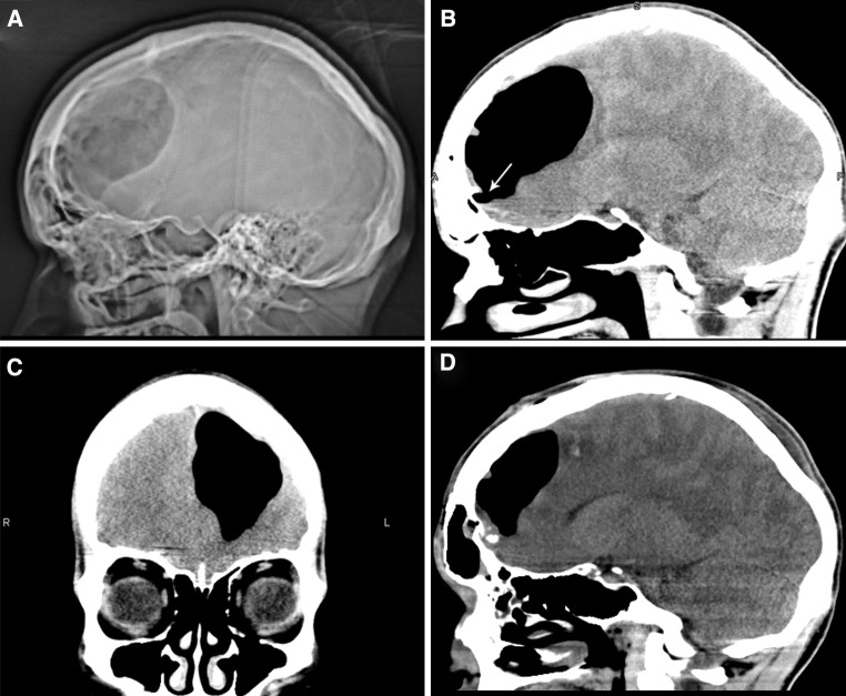
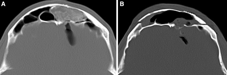
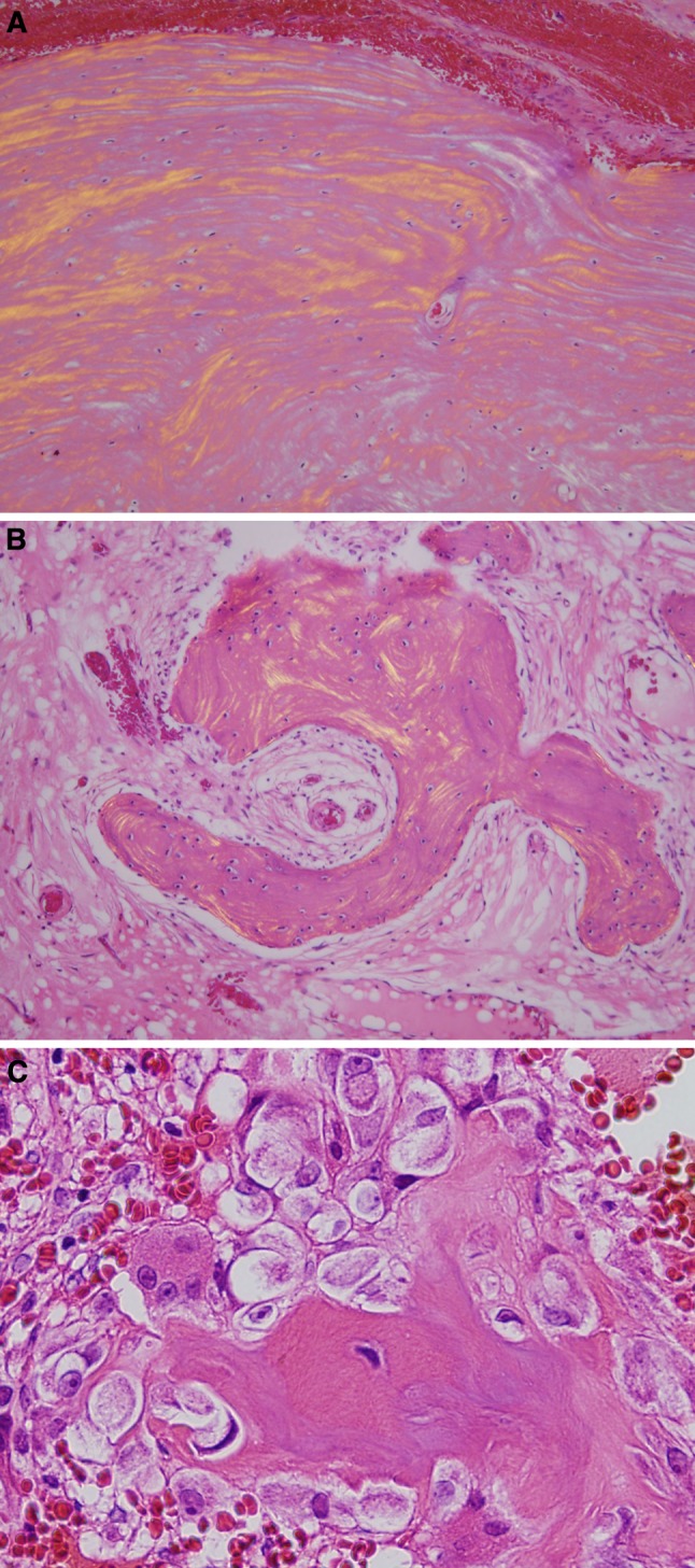
Osteomas of the cranial sinuses are rare, benign bony tumors that can be complicated by the formation of an intracranial pneumatocele. If not treated promptly, a pneumatocele can lead to abscess formation, meningitis, or ventriculitis. In the present case, an intracerebral pneumatocele was formed when an 18 cm(3) osteoma breached the posterior wall of the frontal sinus creating a one-way valve through which air could enter the intracranial cavity. The patient presented after forceful sneezing with nonspecific symptoms of headache, nausea, and vomiting. CT demonstrated a frontal collection of loculated air with mass effect within the left cerebral hemisphere. A partly mineralized mass occupied the left superior nasal ethmoid sinus and left frontal sinus. Of interest pathologically in this case, the tumor had a substantial osteoblastoma-like component. Surgical repair involved frontal craniotomy to remove the osteoma and debride frontal sinus mucosa, plugging the frontal nasal ducts and sinus with fat and bone wax, and dural restoration using an underwater closed drainage system to vent intracranial air and stabilize the patient.
Figures
References
-
- Rappaport J, Attia E. Pneumocephalus in frontal sinus osteoma: a case report. J Otolaryngol. 1994;23:430–436. - PubMed
-
- Herring W. Chapter 21: recognizing abnormalities in bone density. In: Herring W, editor. Learning radiology: recognizing the basics. Philadelphia: Mosby Inc; 2012. pp. 218–229.
-
- Gupta S, Venkatesh SK, Gupta N, et al. Post traumatic intracerebral pneumatocele presenting as CSF rhinorrhea: a case report. Ind J Radiol Imag. 2006;16:735–738. doi: 10.4103/0971-3026.32335. - DOI
Publication types
MeSH terms
LinkOut - more resources
Full Text Sources
Medical
