Review
. 2012 Feb;139(4):641-56.
doi: 10.1242/dev.068353.

Myoblast fusion: lessons from flies and mice

Affiliations
Review

Myoblast fusion: lessons from flies and mice

Susan M Abmayr et al. Development. 2012 Feb.

Abstract

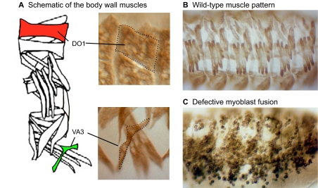
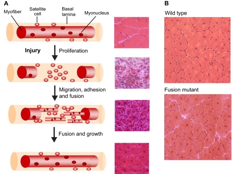
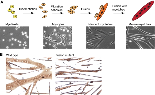
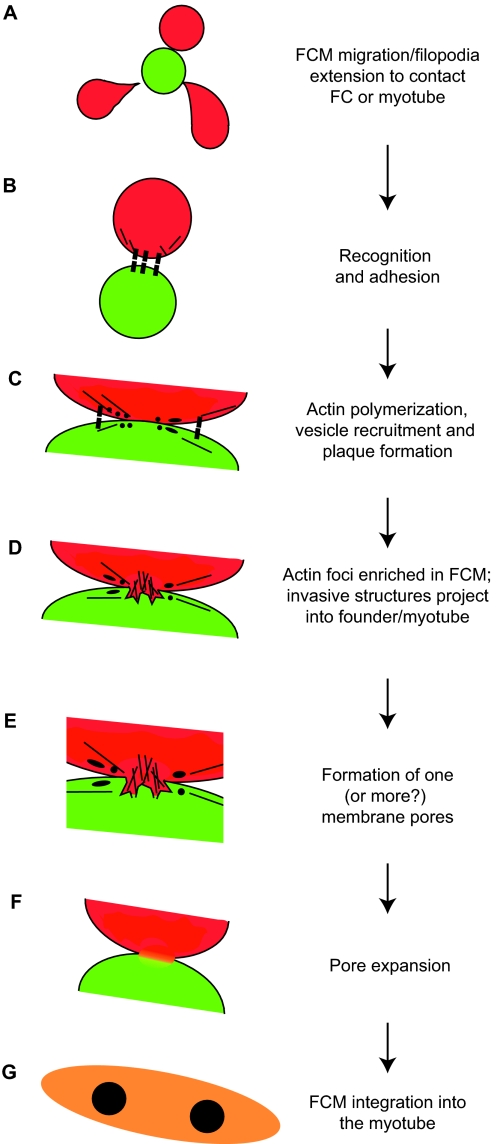
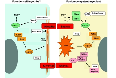
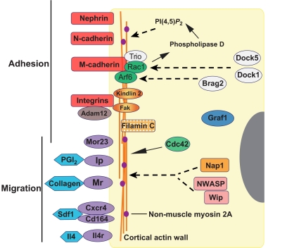
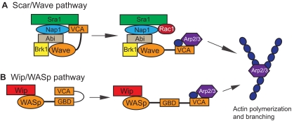
The fusion of myoblasts into multinucleate syncytia plays a fundamental role in muscle function, as it supports the formation of extended sarcomeric arrays, or myofibrils, within a large volume of cytoplasm. Principles learned from the study of myoblast fusion not only enhance our understanding of myogenesis, but also contribute to our perspectives on membrane fusion and cell-cell fusion in a wide array of model organisms and experimental systems. Recent studies have advanced our views of the cell biological processes and crucial proteins that drive myoblast fusion. Here, we provide an overview of myoblast fusion in three model systems that have contributed much to our understanding of these events: the Drosophila embryo; developing and regenerating mouse muscle; and cultured rodent muscle cells.


Figures

Fig. 1.
Fig. 1.
Muscle pattern and myoblast fusion in Drosophila. (A). Left: Schematic of the 30 distinct muscles per abdominal hemisegment in the Drosophila embryo. Highlighted are a small muscle (ventral acute 3; VA3 in green) and a large muscle (dorsal oblique 1; DO1 in red) illustrating the differences in size and shape. Right: High magnification view of VA3 and DO1, as visualized in a wild-type embryo by an antibody directed against muscle myosin. (B,C) The pattern of muscles and myoblasts in wild-type and fusion-defective embryos, as visualized by an antibody against muscle myosin. Multinucleate syncytia are apparent in the wild-type embryo shown in B. Defects in myoblast fusion are easily visible in the mutant embryo (C), highlighting the value of this model system. (B) Reproduced, with permission, from Bour et al. (Bour et al., 1995).
Fig. 2.
Fig. 2.
Muscle regeneration in adult mouse muscle. (A) Each myofiber in adult muscle is surrounded by a basal lamina sheath underneath which lie satellite cells in close apposition to the fiber. In response to injury, segmental necrosis of the myofiber occurs and satellite cells begin to proliferate and form myoblasts. These myoblasts differentiate and then migrate, adhere and fuse with one another to form multiple myotubes within the basal lamina sheath. Myoblasts/myotubes fuse with the stumps of the surviving myofiber and myotubes also fuse with each other to repair the injured myofiber. Regenerated myofibers are easily identified by the presence of centrally located nuclei. For each stage of regeneration in the schematic, a representative mouse muscle section is shown in cross-section and stained with Hematoxylin and Eosin to illustrate the morphological features of the tissue. (B) Cross-sections of regenerated myofibers 14 days after injury from wild-type and mutant mice stained with Hematoxylin and Eosin. Smaller myofibers are observed in the fusion mutant. Note the presence of centrally nucleated myofibers, a hallmark of muscle regeneration.
Fig. 3.
Fig. 3.
Myoblast fusion in cultured muscle cells. (A) Myoblasts proliferate in vitro in medium containing growth factors. To induce myotube formation, growth factors are removed and the majority of myoblasts will terminally differentiate into myocytes, which migrate, adhere and fuse with one another to form small nascent myotubes with few nuclei. Subsequently, nascent myotubes fuse with myocytes and other myotubes to form large mature myotubes with many nuclei. Representative phase contrast photos of mouse muscle cells are shown for each stage. (B) Muscle cells from adult wild-type and mutant mice cultured for 40 hours in vitro and visualized by immunostaining for myosin heavy chain.
Fig. 4.
Fig. 4.
Hypothetical model of myoblast fusion in Drosophila embryos. (A) A fusion-competent myoblast (FCM; red) migrates or extends filopodia to contact a founder cell (FC; green) or, in subsequent rounds of fusion, a syncytial myotube (orange). (B) Cell-surface adhesion molecules (black boxes) mediate recognition and adhesion between cells. (C) Following cell-cell contact, and prior to fusion, electron-dense vesicles (black circles) are recruited to points of contact, possibly through vesicle trafficking mechanisms from the Golgi (not shown). This process may involve actin filaments (black lines). Such vesicles facilitate the fusion process, possibly by delivering fusion-associated components, such as lipids, fusogens or proteases, via targeted exocytosis near or at the sites of fusion. Vesicles may give rise to membrane plaques (black ellipses), which could reflect accumulation of adhesion proteins or other fusion machinery. (D) Actin accumulates in the FCM, forming a large F-actin-based protrusion that pushes into the founder cell. A thin sheath of actin is present in the founder cell (not shown). (E) One, or more, fusion pores form to allow mixing of cytoplasmic contents. (F) Expansion of the fusion pore(s) and elimination of membrane separating the cells. (G) The FCM is absorbed into the myotube, and the resulting syncytium continues additional rounds of fusion as needed.
Fig. 5.
Fig. 5.
Genes and pathways associated with myoblast fusion in Drosophila. The indicated genes and pathways correspond to those, as discussed in the text, that appear to function in founder cells/myotubes and fusion-competent myoblasts of Drosophila embryos. The represented proteins include those for which a role in fusion has been shown experimentally. Generally, these comprise components of the Rac1, Scar and WASp pathways and their regulators, cell-adhesion molecules, and essential proteins such as Sing, for which a mechanistic role remains to be elucidated. In most instances, demonstration of an involvement in myoblast fusion has been established by direct loss-of-function studies experimentally. For some proteins, a role is inferred by biochemical interaction with known fusion proteins. Relationships indicated by broken lines are based on protein functions in other tissues, but have not yet been established in this system. Arp2/3, Actin-related protein 2/3; Ants, Antisocial; Arf6, ADP ribosylation factor 6; Blow, Blown Fuse; Crk, CT10 regulator of Kinase; Elmo, Engulfment and cell motility protein; GEF, Guanine nucleotide exchange factor; Hbs, Hibris; Kirre, Kin-of IrreC; Mbc, Myoblast city; PIP3, Phosphatidylinositol (3,4,5) triphosphate; Rac1, Ras-related C3 botulinum toxin substrate 1; Rst, Roughest; Sltr, Solitary; Rols7, Rolling pebbles isoform 7; Scar, Suppressor of cAMP receptor; Sing, Singles-bar; Sns, Sticks and stones; Vrp1, Verprolin 1; WASp, Wiscott-Aldrich syndrome protein; Wip, Drosophila WASp-interacting protein.
Fig. 6.
Fig. 6.
Genes and pathways associated with myoblast fusion in mice. The indicated proteins and pathways correspond to those, as discussed in the text, for which a role in fusion has been shown experimentally. Generally, these comprise cell-adhesion molecules and their associated adaptors, secreted molecules and their receptors that control cell migration, and molecules that signal to the actin cytoskeleton. For the most part, roles for these proteins have been identified through loss-of-function studies. Relationships indicated by broken lines are based on studies in other tissues, but have not yet been established in this system. Arf6, ADP ribosylation factor 6; Brag2, brefeldin A-resistant Arf GEF; CD164, cluster of differentiation 164; Cdc42, cell division cycle 42; Cxcr4, chemokine (C-X-C motif) receptor 4; Dock1 and Dock 5, dedicator of cytokinesis 1 and 5; Fak, focal adhesion kinase; Graf1, GTPase regulator associated with focal adhesion kinase; Il4, interleukin 4; Il4r, interleukin 4 receptor; Ip, prostacyclin receptor; Mr, mannose receptor; Mor23, mouse odorant receptor 23; Nap1, Nck-associated protein; NWASP, neural Wiskott-Aldrich syndrome protein; PGI2, prostacyclin; PI(4,5)P2, phosphatidylinositol 4,5-bisphosphate; Rac1, Ras-related C3 botulinum toxin substrate 1; SDF1, stromal-derived factor 1; Wip, WASp-interacting protein.
Fig. 7.
Fig. 7.
Protrusion of FCM in developing myotube. (A) A single confocal slice through the middle of a primary myoblast and its associated myofiber. Kirre protein (turquoise) and Sns protein (green) are highly enriched at the point of protrusion. Actin (red) is highly restricted to the fusion-competent myoblast, although faint actin fibers are visible in the myotube. A dotted line outlines the surface of the myotube and fusion-competent myoblast. Reproduced, with permission, from Haralalka et al. (Haralalka et al., 2011). (B) Schematic of the image shown in A.
None

References

    1. Abramovici H., Gee S. H. (2007). Morphological changes and spatial regulation of diacylglycerol kinase-zeta, syntrophins, and Rac1 during myoblast fusion. Cell Motil. Cytoskel. 64, 549–567 - PubMed
    1. Albiges-Rizo C., Destaing O., Fourcade B., Planus E., Block M. R. (2009). Actin machinery and mechanosensitivity in invadopodia, podosomes and focal adhesions. J. Cell Sci. 122, 3037–3049 - PMC - PubMed
    1. Anton I. M., Jones G. E., Wandosell F., Geha R., Ramesh N. (2007). WASP-interacting protein (WIP): working in polymerisation and much more. Trends Cell Biol. 17, 555–562 - PubMed
    1. Arnold L., Henry A., Poron F., Baba-Amer Y., van Rooijen N., Plonquet A., Gherardi R. K., Chazaud B. (2007). Inflammatory monocytes recruited after skeletal muscle injury switch into antiinflammatory macrophages to support myogenesis. J. Exp. Med. 204, 1057–1069 - PMC - PubMed
    1. Artero R. D., Castanon I., Baylies M. K. (2001). The immunoglobulin-like protein Hibris functions as a dose-dependent regulator of myoblast fusion and is differentially controlled by Ras and Notch signaling. Development 128, 4251–4264 - PubMed

Publication types

MeSH terms

Substances

LinkOut - more resources