

Adhesive forces in embryonic stem cell cultures
- PMID: 22274712
- PMCID: PMC3277780
- DOI: 10.4161/cam.5.6.18270
Adhesive forces in embryonic stem cell cultures
Abstract
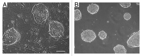
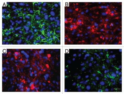
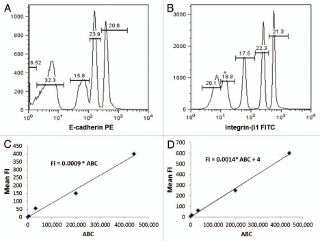
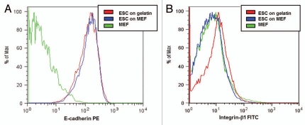
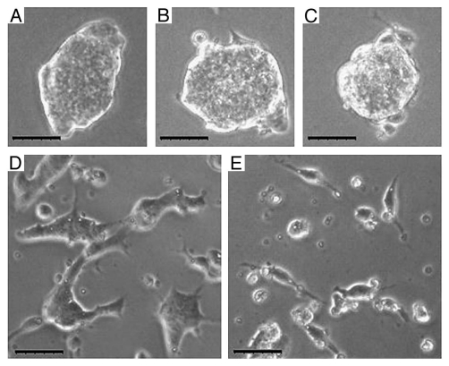
Most cell culture systems grow and spread as contact-inhibited monolayers on flat culture dishes, but the embryonic stem cell (ESC) is one of the cell phenotypes that prefer to self-organize as tightly packed three-dimensional (3D) colonies. ESC also readily form 3D cell aggregates, called embryoid bodies (EB) that partially mimic the spatial and temporal processes of the developing embryo. Here, the rationale for ESC aggregatation, rather than "spreading" on gelatin-coated or mouse embryonic fibroblast (MEF)-coated dishes, is examined through the quantification of the expression levels of adhesion molecules on ESC and the calculation of the adhesive forces on ESC. Modeling each ESC as a dodecahedron, the adhesive force for each ESC-ESC binding was found to be 9.1 x 10(5) pN, whereas, the adhesive force for ESC-MEF binding was found to be an order of magnitude smaller at 7.9 x 10(4) pN. We also show that E-cadherin is the dominating molecule in the ESC-ESC adhesion and blocking E-cadherin leads to a significant reduction in colony formation. Here, we mathematically describe the preference for ESC to self-assemble into ESC-ESC aggregates and 3D colonies, rather than to bind and spread on gelatin or MEF-coated dishes, and have shown that these interactions are predominantly due to E-cadherin expression on ESC.
Figures
Similar articles
-
Differential adhesion molecule expression during murine embryonic stem cell commitment to the hematopoietic and endothelial lineages.PLoS One. 2011;6(9):e23810. doi: 10.1371/journal.pone.0023810. Epub 2011 Sep 6. PLoS One. 2011. PMID: 21909405 Free PMC article.
-
The primitive endoderm segregates from the epiblast in β1 integrin-deficient early mouse embryos.Mol Cell Biol. 2014 Feb;34(3):560-72. doi: 10.1128/MCB.00937-13. Epub 2013 Nov 25. Mol Cell Biol. 2014. PMID: 24277939 Free PMC article.
-
Embryonic stem cells growing in 3-dimensions shift from reliance on the substrate to each other for mechanical support.J Biomech. 2015 Jul 16;48(10):1777-81. doi: 10.1016/j.jbiomech.2015.05.009. Epub 2015 May 21. J Biomech. 2015. PMID: 26050958
-
Role of cell-cell adhesion complexes in embryonic stem cell biology.J Cell Sci. 2014 Jun 15;127(Pt 12):2603-13. doi: 10.1242/jcs.146720. J Cell Sci. 2014. PMID: 24931943 Review.
-
Cell Mechanics in Embryoid Bodies.Cells. 2020 Oct 11;9(10):2270. doi: 10.3390/cells9102270. Cells. 2020. PMID: 33050550 Free PMC article. Review.
Cited by
-
The Sall2 transcription factor promotes cell migration regulating focal adhesion turnover and integrin β1 expression.Front Cell Dev Biol. 2022 Nov 9;10:1031262. doi: 10.3389/fcell.2022.1031262. eCollection 2022. Front Cell Dev Biol. 2022. PMID: 36438565 Free PMC article.
-
A 3D magnetic tissue stretcher for remote mechanical control of embryonic stem cell differentiation.Nat Commun. 2017 Sep 12;8(1):400. doi: 10.1038/s41467-017-00543-2. Nat Commun. 2017. PMID: 28900152 Free PMC article.
-
Covariation of Pluripotency Markers and Biomechanical Properties in Mouse Embryonic Stem Cells.Front Cell Dev Biol. 2022 May 16;10:858884. doi: 10.3389/fcell.2022.858884. eCollection 2022. Front Cell Dev Biol. 2022. PMID: 35652102 Free PMC article.
References
-
- Larue L. A role for cadherins in tissue formation. Development. 1996;122:3185–3194. - PubMed
Publication types
MeSH terms
Substances
Grants and funding
LinkOut - more resources
Full Text Sources