

Can mesenchymal cells undergo collective cell migration? The case of the neural crest
- PMID: 22274714
- PMCID: PMC3277782
- DOI: 10.4161/cam.5.6.18623
Can mesenchymal cells undergo collective cell migration? The case of the neural crest
Abstract
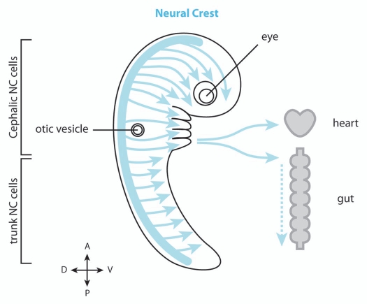
Cell migration is critical for proper development of the embryo and is also used by many cell types to perform their physiological function. For instance, cell migration is essential for immune cells to monitor the body and for epithelial cells to heal a wound whereas, in cancer cells, acquisition of migratory capabilities is a critical step towards malignancy. Migratory cells are often categorized into two groups: mesenchymal cells, produced by an epithelium-to-mesenchyme transition, that undergo solitary migration and epithelial-like cells which migrate collectively. However, on some occasions, mesenchymal cells may travel in large, dense groups and exhibit key features of collectively migrating cells such as coordination and cooperation. Here, using data published on Neural Crest cells, a highly invasive mesenchymal cell population that extensively migrate throughout the embryo, we explore the idea that other mesenchymal cells, including cancer cells, might be able to undergo collective cell migration under certain conditions and discuss how they could do so.
Figures
Similar articles
-
Embryonic cell-cell adhesion: a key player in collective neural crest migration.Curr Top Dev Biol. 2015;112:301-23. doi: 10.1016/bs.ctdb.2014.11.023. Epub 2015 Feb 12. Curr Top Dev Biol. 2015. PMID: 25733144 Review.
-
Cadherins in collective cell migration of mesenchymal cells.Curr Opin Cell Biol. 2012 Oct;24(5):677-84. doi: 10.1016/j.ceb.2012.08.002. Epub 2012 Sep 1. Curr Opin Cell Biol. 2012. PMID: 22944726 Free PMC article. Review.
-
Mechanisms of Neural Crest Migration.Annu Rev Genet. 2018 Nov 23;52:43-63. doi: 10.1146/annurev-genet-120417-031559. Annu Rev Genet. 2018. PMID: 30476447 Review.
-
Tissue stiffening coordinates morphogenesis by triggering collective cell migration in vivo.Nature. 2018 Feb 22;554(7693):523-527. doi: 10.1038/nature25742. Epub 2018 Feb 14. Nature. 2018. PMID: 29443958 Free PMC article.
-
Neural crest and mesoderm lineage-dependent gene expression in orofacial development.Differentiation. 2007 Jun;75(5):463-77. doi: 10.1111/j.1432-0436.2006.00145.x. Epub 2007 Feb 5. Differentiation. 2007. PMID: 17286603
Cited by
-
Viscoelasticity, Like Forces, Plays a Role in Mechanotransduction.Front Cell Dev Biol. 2022 Feb 9;10:789841. doi: 10.3389/fcell.2022.789841. eCollection 2022. Front Cell Dev Biol. 2022. PMID: 35223831 Free PMC article. Review.
-
The plasticity of pancreatic cancer stem cells: implications in therapeutic resistance.Cancer Metastasis Rev. 2021 Sep;40(3):691-720. doi: 10.1007/s10555-021-09979-x. Epub 2021 Aug 28. Cancer Metastasis Rev. 2021. PMID: 34453639 Free PMC article. Review.
-
Rho GTPase signalling in cell migration.Curr Opin Cell Biol. 2015 Oct;36:103-12. doi: 10.1016/j.ceb.2015.08.005. Epub 2015 Sep 10. Curr Opin Cell Biol. 2015. PMID: 26363959 Free PMC article. Review.
-
Androgen-regulated MafB drives cell migration via MMP11-dependent extracellular matrix remodeling in mice.iScience. 2022 Nov 16;25(12):105609. doi: 10.1016/j.isci.2022.105609. eCollection 2022 Dec 22. iScience. 2022. PMID: 36465133 Free PMC article.
-
The extracellular matrix in development.Development. 2020 May 28;147(10):dev175596. doi: 10.1242/dev.175596. Development. 2020. PMID: 32467294 Free PMC article. Review.
References
-
- Hall B. The neural crest and neural crest cells in vertebrate development and evolution. New York: Springer; 2008.
-
- Le Douarin N, Kalcheim C. The neural crest. Cambridge UK; New York NY, USA: Cambridge University Press; 1999.
Publication types
MeSH terms
Grants and funding
LinkOut - more resources
Full Text Sources