. 2012 Apr;17(4):421-34.
doi: 10.1177/1087057111433843. Epub 2012 Jan 24.

Identification and mechanistic studies of a novel ubiquitin E1 inhibitor

Affiliations

Identification and mechanistic studies of a novel ubiquitin E1 inhibitor

Dana Ungermannova et al. J Biomol Screen. 2012 Apr.

Abstract

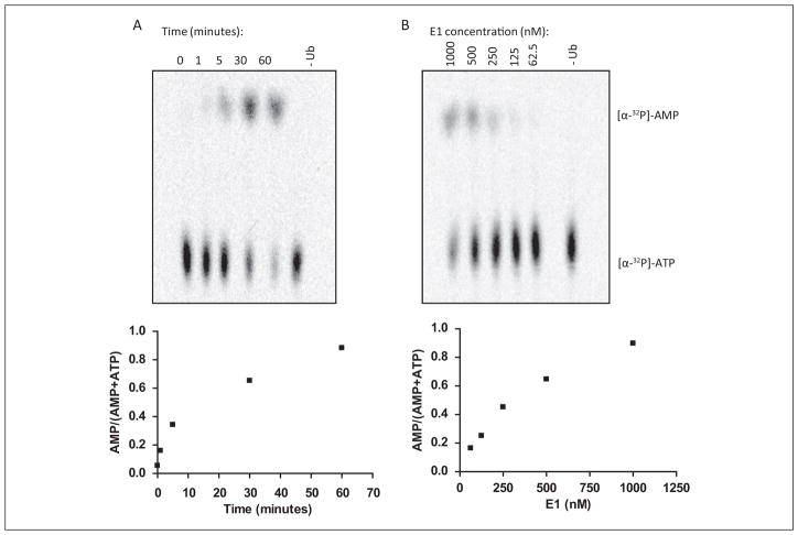
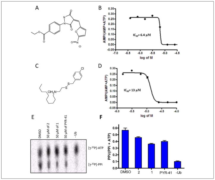
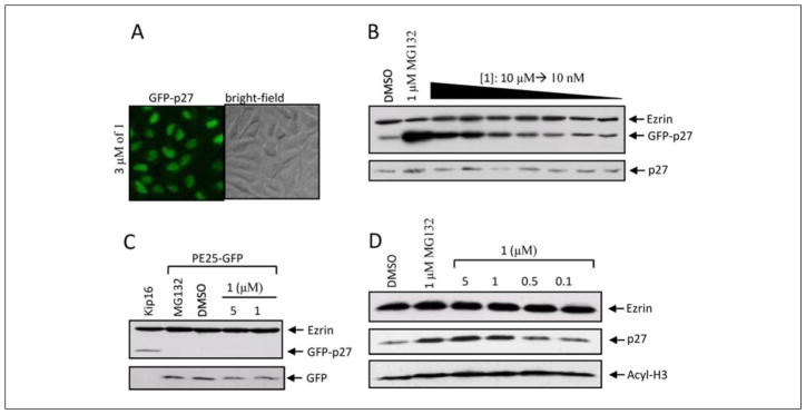
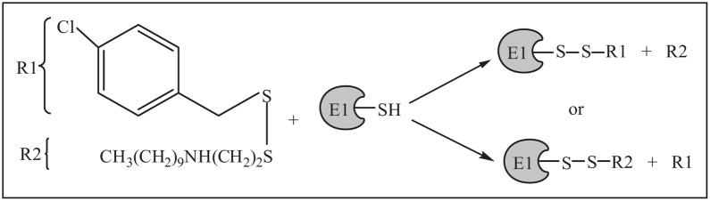
Protein degradation via the ubiquitin-proteasome pathway is important for a diverse number of cellular processes ranging from cell signaling to development. Disruption of the ubiquitin pathway occurs in a variety of human diseases, including several cancers and neurological disorders. Excessive proteolysis of tumor suppressor proteins, such as p27, occurs in numerous aggressive human tumors. To discover small-molecule inhibitors that potentially prevent p27 degradation, we developed a series of screening assays, including a cell-based screen of a small-molecule compound library and two novel nucleotide exchange assays. Several small-molecule inhibitors, including NSC624206, were identified and subsequently verified to prevent p27 ubiquitination in vitro. The mechanism of NSC624206 inhibition of p27 ubiquitination was further unraveled using the nucleotide exchange assays and shown to be due to antagonizing ubiquitin activating enzyme (E1). We determined that NSC624206 and PYR-41, a recently reported inhibitor of ubiquitin E1, specifically block ubiquitin-thioester formation but have no effect on ubiquitin adenylation. These studies reveal a novel E1 inhibitor that targets a specific step of the E1 activation reaction. NSC624206 could, therefore, be potentially useful for the control of excessive ubiquitin-mediated proteolysis in vivo.


Conflict of interest statement

Declaration of Conflicting Interests

The authors declared no potential conflicts of interest with respect to the research, authorship, and/or publication of this article.

Figures

Figure 1
Figure 1
Identification of inhibitors of p27 proteolysis from a library of small molecules using a cell-based assay. (A) Cell-based assay to test a library of compounds with a high-throughput screen (HTS). A HTS was developed to detect compounds that increased the fluorescence of a green fluorescent protein (GFP)–p27 reporter. Kip16 cells were seeded at a density of 40 000 cells/well in a 96-well plate. DMSO was added to column 1 to establish a background signal, 1 μM MG132 was added to column 12 to establish a maximum signal (positive control), and the remaining 80 wells received 3 μM of the specific compound. Plates were read on a Safire II plate reader, and the results were plotted as percentage of fluorescence relative to MG132 using Microsoft Excel. The results presented here are from National Cancer Institute (NCI) plate 4149, where wells 6E and 6G are considered possible hits given that the measured signal for both compounds was comparable to that of MG132. (B) The dot plot of library screen results. In total, 3161 compounds (40 plates) were tested in the cell-based assay. The results were quantified and compiled as the number of compounds found for different fluorescence readings using statistical software Minitab 15 (Minitab, State College, PA). Each data point represents up to 20 compounds (see Suppl. Fig. S1D for histograms of the frequency distribution of the signals from compounds, DMSO, and MG132 control signals). (C) Fluorescent and corresponding bright-field images of Kip16 cells. Addition of MG132 (1 μM) blocks the degradation of GFP-p27, whereas DMSO has no effect on the reporter protein stabilization.
Figure 2
Figure 2
Secondary screening of compounds identified in the cell-based assay. (A) Reconstitution of the p27 ubiquitination reaction in the presence of 300 μM of the hit compounds. The reactions were performed as described in Materials and Methods. Four compounds (lanes 8, 13, 15, 19) reduced the levels of polyubiquitinated p27 when compared with the vehicle (lanes 2 and 12). (B) Structures of the hit compounds directly adapted from http://dtp.cancer.gov. (C) Chemical structures of NSC624206 and its analog prepared synthetically. 1 is an asymmetric disulfide compound with the same structure as NSC624206. 2 is a derivative of 1 where the disulfide was replaced with a sulfide to explore the importance of the disulfide linkage.
Figure 3
Figure 3
1 selectively inhibits ubiquitin E1 enzymes in vitro. (A) Thioester assay of E1/E2 activity using fluorescein ubiquitin (Ub-F). Thioester bond formation between E1 and Ub-F is adenosine triphosphate (ATP) dependent (lane 2 vs. lane 1). Upon addition of purified E2 (His-Cdc34), an E2-ubiquitin adduct is observed with a concomitant reduction of the E1 thioester (lane 3). As expected for thioester-bonded adducts, high concentrations of dithiothreitol (DTT) eliminate ubiquitin from E1 and E2 as shown in lane 5 (100 mM DTT), lane 6 (10 mM DTT), and lane 7 (1 mM DTT). (B) 1, but not 2, inhibits the transfer of ubiquitin from E1 to E2. In total, 50 nM E1 was incubated with decreasing concentrations of 1 or 2 (50, 25, and 1 μM—lanes 2, 3, and 4, respectively) for 10 min at room temperature followed by the addition of a cocktail containing ATP, E2, and Ub-F. This was allowed to react for 5 min. The reactions were resolved by sodium dodecyl sulfate polyacrylamide gel electrophoresis (SDS-PAGE) under nonreducing conditions. Although 2 does not interfere with the attachment of ubiquitin onto E1/E2, the thioester adducts disappear in the presence of 50 and 25 μM of 1. DMSO has no effect on the formation of the thioester linkage (lane 1). (C) 1 inhibits transfer onto E1 in a concentration-dependent manner with IC50 of 9 μM. In total, 50 nM of E1 was incubated with decreasing concentrations of 1 (50 to 1 μM), and the results were analyzed using ImageJ (National Institutes of Health, Bethesda, MD), and IC50 was extracted using the GraphPad program (GraphPad Software, San Diego, CA).
Figure 4
Figure 4
Reaction steps within E1 activation tested by [α-32P]-AMP:[α-32P]-ATP exchange assay. AMP, adenosine monophosphate; ATP, adenosine triphosphate.
Figure 5
Figure 5
Development of nucleotide exchange assay to monitor ubiquitin-dependent ATP:AMP exchange during E1 activation. (A) Time course of ATP:AMP exchange assay. In total, 1 μM E1, 100 μM cold ATP, 1 μM hot ATP, 2 mM cold AMP, and 500 μM PPi were mixed together in magnesium2+-containing thioester assay buffer. The reaction was initiated by adding ubiquitin to a final concentration of 5 μM and incubated at 37 °C. The reactions were quenched at the indicated times with EDTA and run on a thin-layer chromatography (TLC) plate in potassium phosphate buffer for 15 min followed by autoradiography. The lower panel shows the corresponding quantification of the time course where the ratio of the two nucleotides was determined for each time point. (B) E1 concentration dependence. In total, 1 μM to 62.5 nM E1 was added to the exchange reaction for 10 min at 37 °C. The reactions were developed as above. The amount of labeled AMP produced from ATP was quantified and plotted for each E1 concentration. AMP, adenosine monophosphate; ATP, adenosine triphosphate; PPi, pyrophosphate.
Figure 6
Figure 6
Determination of IC50 for PYR-41 and 1 using the ATP:AMP exchange assay. (A) Structure of PYR-41. (B) PYR-41 inhibits E1 in a concentration-dependent fashion. PYR-41 was serially diluted (50 to 0.39 μM) and incubated with 150 nM E1 at room temperature for 10 min, and then the thioester reaction mixture was added with ubiquitin to initiate the nucleotide exchange. All reactions were halted with addition of EDTA after a 10-min incubation at 37 °C and resolved using thin-layer chromatography (TLC) plates followed by PhosphorImager (GE Healthcare, Piscataway, NJ) analysis (see Suppl. Fig. S4A for TLC plate image). ImageJ was used to quantify the amounts of both nucleotides, and their ratio was plotted against a logarithmic scale of PYR-41 concentration using the GraphPad program (GraphPad Software, San Diego, CA), which was also used to calculate IC50. The IC50 value is the average from one experiment that was run twice on the same TLC plate. (C) Structure of 1 for reference. (D) 1 inhibits E1 in a concentration-dependent manner. E1 was incubated with decreasing concentrations of 1 (100 to 1.5 μM, developed and quantified as described in (B). See Supplementary Figure S4B for the TLC plate image. (E) 1 and PYR-41 do not affect the production of pyrophosphate (PPi) during E1 adenylation as measured by the [32P]-PPi:[γ-32P]-ATP exchange assay. In total, 150 nM E1, 100 μM cold ATP, 1 μM [γ-32P]-ATP, 2 mM cold AMP, and 500 μM PPi were mixed together in an Mg2+-containing thioester assay buffer. The reaction was initiated by adding ubiquitin to a final concentration of 5 μM and incubated at 37 °C. The reactions were quenched at indicated times with EDTA and run on a TLC plate in potassium phosphate buffer for 15 min followed by autoradiography. Incubation of E1 with DMSO generated the most pyrophosphate since the E1 activating reaction proceeded to completion. Less PPi was released in the presence of 1 and PYR-41 since the reaction stopped at step 2 and no PPi was generated without ubiquitin. (F) Quantification of the [32P]-PPi:[γ-32P]-ATP exchange assay. AMP, adenosine monophosphate; ATP, adenosine triphosphate.
Figure 7
Figure 7
1 induces accumulation of p27 in cells. (A) Fluorescence and corresponding bright-field images of Kip16 cells; 3 μM 1 uniformly stabilizes green fluorescent protein (GFP)–p27. (B) 1 treatment induces the expression of GFP-p27 in a dose-dependent manner but does not affect endogenous p27. Kip16 cells were treated with 1 at concentrations ranging from 10 μM to 10 nM for 24 h, and the amount of total p27 was determined by Western blotting using an anti-p27 antibody. A total of 1 μM MG132, which significantly upregulates GFP-p27, was used as a positive control. (C) 1 does not alter the expression of GFP alone. PE25 cells (mink lung cells stably expressing GFP) were treated with DMSO or 1 μM MG132 or 5 or 1 μM 1 for 24 h. Then, 50 μg of cell lysates was subjected to immunoblot analysis with an anti-GFP antibody. DMSO-treated Kip16 cells were used to show the GFP antibody specificity. (D) 1 increases p27 in a dose-dependent manner in HepG2 liver cancer cells. HepG2 cells were incubated with MG132 or 1 as indicated for 24 h, and the cell extracts were resolved by sodium dodecyl sulfate polyacrylamide gel electrophoresis (SDS-PAGE) followed by detection with indicated antibodies. In every Western blot presented in this figure, the expression of ezrin was used to show equal loading.
Figure 8
Figure 8
Proposed reaction mechanism of 1 with E1. Reaction of disulfide compound 1 with the active site cysteine of E1 causes the release of p-chlorobenzyl thiol (R1) and decylamino ethane thiol-E1 (R2), which results in inhibition of the enzyme. It is also possible that during the thiol-disulfide exchange reaction, the R groups are interchanged.

Similar articles

Cited by

References

    1. Hershko A, Ciechanover A. Mechanisms of Intracellular Protein Breakdown. Annu Rev Biochem. 1982;51:335–364. - PubMed
    1. Dikic I, Wakatsuki S, Walters KJ. Ubiquitin-binding Domains—From Structures to Functions. Nat Rev Mol Cell Biol. 2009;10:659–671. - PMC - PubMed
    1. Kane RC, Bross PF, Farrell AT, Pazdur R. Velcade: U.S. FDA Approval for the Treatment of Multiple Myeloma Progressing on Prior Therapy. Oncologist. 2003;8:508–513. - PubMed
    1. San Miguel JF, Schlag R, Khuageva NK, Dimopoulos MA, Shpilberg O, Kropff M, Spicka I, Petrucci MT, Palumbo A, Samoilova OS, et al. Bortezomib Plus Melphalan and Prednisone for Initial Treatment of Multiple Myeloma. N Engl J Med. 2008;359:906–917. - PubMed
    1. Pagano M, Tam SW, Theodoras AM, Beer-Romero P, Del Sal G, Chau V, Yew PR, Draetta GF, Rolfe M. Role of the Ubiquitin-Proteasome Pathway in Regulating Abundance of the Cyclin-Dependent Kinase Inhibitor p27. Science. 1995;269:682–685. - PubMed

Publication types

MeSH terms