An in vivo selection method to optimize trans-splicing ribozymes
- PMID: 22274958
- PMCID: PMC3285944
- DOI: 10.1261/rna.028472.111
An in vivo selection method to optimize trans-splicing ribozymes
Abstract
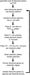
Group I intron ribozymes can repair mutated mRNAs by replacing the 3'-terminal portion of the mRNA with their own 3'-exon. This trans-splicing reaction has the potential to treat genetic disorders and to selectively kill cancer cells or virus-infected cells. However, these ribozymes have not yet been used in therapy, partially due to a low in vivo trans-splicing efficiency. Previous strategies to improve the trans-splicing efficiencies focused on designing and testing individual ribozyme constructs. Here we describe a method that selects the most efficient ribozymes from millions of ribozyme variants. This method uses an in vivo rescue assay where the mRNA of an inactivated antibiotic resistance gene is repaired by trans-splicing group I intron ribozymes. Bacterial cells that express efficient trans-splicing ribozymes are able to grow on medium containing the antibiotic chloramphenicol. We randomized a 5'-terminal sequence of the Tetrahymena thermophila group I intron and screened a library with 9 × 10⁶ ribozyme variants for the best trans-splicing activity. The resulting ribozymes showed increased trans-splicing efficiency and help the design of efficient trans-splicing ribozymes for different sequence contexts. This in vivo selection method can now be used to optimize any sequence in trans-splicing ribozymes.
Figures


Similar articles
-
Design and Experimental Evolution of trans-Splicing Group I Intron Ribozymes.Molecules. 2017 Jan 2;22(1):75. doi: 10.3390/molecules22010075. Molecules. 2017. PMID: 28045452 Free PMC article. Review.
-
Trans-splicing with the group I intron ribozyme from Azoarcus.RNA. 2014 Feb;20(2):202-13. doi: 10.1261/rna.041012.113. Epub 2013 Dec 16. RNA. 2014. PMID: 24344321 Free PMC article.
-
Computational prediction of efficient splice sites for trans-splicing ribozymes.RNA. 2012 Mar;18(3):590-602. doi: 10.1261/rna.029884.111. Epub 2012 Jan 24. RNA. 2012. PMID: 22274956 Free PMC article.
-
RNA reprogramming of alpha-mannosidase mRNA sequences in vitro by myxomycete group IC1 and IE ribozymes.FEBS J. 2006 Jun;273(12):2789-800. doi: 10.1111/j.1742-4658.2006.05295.x. FEBS J. 2006. PMID: 16817905
-
Group I Intron-Based Therapeutics Through Trans-Splicing Reaction.Prog Mol Biol Transl Sci. 2018;159:79-100. doi: 10.1016/bs.pmbts.2018.07.001. Epub 2018 Aug 9. Prog Mol Biol Transl Sci. 2018. PMID: 30340790 Review.
Cited by
-
Design and Experimental Evolution of trans-Splicing Group I Intron Ribozymes.Molecules. 2017 Jan 2;22(1):75. doi: 10.3390/molecules22010075. Molecules. 2017. PMID: 28045452 Free PMC article. Review.
-
Site-Selective RNA Splicing Nanozyme: DNAzyme and RtcB Conjugates on a Gold Nanoparticle.ACS Chem Biol. 2018 Jan 19;13(1):215-224. doi: 10.1021/acschembio.7b00437. Epub 2017 Dec 19. ACS Chem Biol. 2018. PMID: 29155548 Free PMC article.
-
Efficient circular RNA synthesis for potent rolling circle translation.Nat Biomed Eng. 2025 Jul;9(7):1062-1074. doi: 10.1038/s41551-024-01306-3. Epub 2024 Dec 13. Nat Biomed Eng. 2025. PMID: 39672985 Free PMC article.
-
Increased efficiency of evolved group I intron spliceozymes by decreased side product formation.RNA. 2015 Aug;21(8):1480-9. doi: 10.1261/rna.051888.115. Epub 2015 Jun 23. RNA. 2015. PMID: 26106216 Free PMC article.
-
Low selection pressure aids the evolution of cooperative ribozyme mutations in cells.J Biol Chem. 2013 Nov 15;288(46):33096-106. doi: 10.1074/jbc.M113.511469. Epub 2013 Oct 2. J Biol Chem. 2013. PMID: 24089519 Free PMC article.
References
-
- Beaudry AA, Joyce GF 1992. Directed evolution of an RNA enzyme. Science 257: 635–641 - PubMed
Publication types
MeSH terms
Substances
Grants and funding
LinkOut - more resources
Full Text Sources
Other Literature Sources
