

Optical coherence tomography captures rapid hemodynamic responses to acute hypoxia in the cardiovascular system of early embryos
- PMID: 22275053
- PMCID: PMC3295592
- DOI: 10.1002/dvdy.23727
Optical coherence tomography captures rapid hemodynamic responses to acute hypoxia in the cardiovascular system of early embryos
Abstract
Background: The trajectory to heart defects may start in tubular and looping heart stages when detailed analysis of form and function is difficult by currently available methods. We used a novel method, Doppler optical coherence tomography (OCT), to follow changes in cardiovascular function in quail embryos during acute hypoxic stress. Chronic fetal hypoxia is a known risk factor for congenital heart diseases (CHDs). Decreased fetal heart rates during maternal obstructive sleep apnea suggest that studying fetal heart responses under acute hypoxia is warranted.
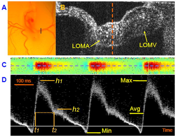
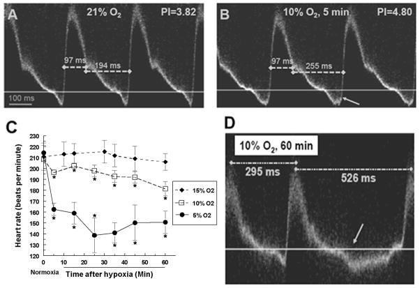
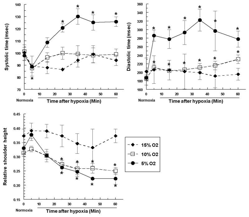
Results: We captured responses to hypoxia at the critical looping heart stages. Doppler OCT revealed detailed vitelline arterial pulsed Doppler waveforms. Embryos tolerated 1 hr of hypoxia (5%, 10%, or 15% O(2) ), but exhibited changes including decreased systolic and increased diastolic duration in 5 min. After 5 min, slower heart rates, arrhythmic events and an increase in retrograde blood flow were observed. These changes suggested slower filling of the heart, which was confirmed by four-dimensional Doppler imaging of the heart itself.
Conclusions: Doppler OCT is well suited for rapid noninvasive screening for functional changes in avian embryos under near physiological conditions. Analysis of the accessible vitelline artery sensitively reflected changes in heart function and can be used for rapid screening. Acute hypoxia caused rapid hemodynamic changes in looping hearts and may be a concern for increased CHD risk.
Copyright © 2012 Wiley Periodicals, Inc.
Figures

Similar articles
-
Cardiac neural crest ablation results in early endocardial cushion and hemodynamic flow abnormalities.Am J Physiol Heart Circ Physiol. 2016 Nov 1;311(5):H1150-H1159. doi: 10.1152/ajpheart.00188.2016. Epub 2016 Aug 19. Am J Physiol Heart Circ Physiol. 2016. PMID: 27542407 Free PMC article.
-
Increased regurgitant flow causes endocardial cushion defects in an avian embryonic model of congenital heart disease.Congenit Heart Dis. 2017 May;12(3):322-331. doi: 10.1111/chd.12443. Epub 2017 Feb 17. Congenit Heart Dis. 2017. PMID: 28211263 Free PMC article.
-
Ethanol exposure alters early cardiac function in the looping heart: a mechanism for congenital heart defects?Am J Physiol Heart Circ Physiol. 2014 Feb;306(3):H414-21. doi: 10.1152/ajpheart.00600.2013. Epub 2013 Nov 22. Am J Physiol Heart Circ Physiol. 2014. PMID: 24271490 Free PMC article.
-
[Congenital heart disease and acquired valvular lesions in pregnancy].Herz. 2003 May;28(3):227-39. doi: 10.1007/s00059-003-2467-y. Herz. 2003. PMID: 12756480 Review. German.
-
Cardiovascular developmental insights from embryos.Ann N Y Acad Sci. 2007 Apr;1101:377-88. doi: 10.1196/annals.1389.012. Epub 2007 Feb 15. Ann N Y Acad Sci. 2007. PMID: 17303838 Review.
Cited by
-
Speckle variance optical coherence tomography of blood flow in the beating mouse embryonic heart.J Biophotonics. 2017 May;10(5):735-743. doi: 10.1002/jbio.201600293. Epub 2017 Apr 18. J Biophotonics. 2017. PMID: 28417585 Free PMC article.
-
In vivo functional imaging of blood flow and wall strain rate in outflow tract of embryonic chick heart using ultrafast spectral domain optical coherence tomography.J Biomed Opt. 2012 Sep;17(9):96006-1. doi: 10.1117/1.JBO.17.9.096006. J Biomed Opt. 2012. PMID: 23085907 Free PMC article.
-
Quantitative optical imaging of vascular response in vivo in a model of peripheral arterial disease.Am J Physiol Heart Circ Physiol. 2013 Oct 15;305(8):H1168-80. doi: 10.1152/ajpheart.00362.2013. Epub 2013 Aug 16. Am J Physiol Heart Circ Physiol. 2013. PMID: 23955718 Free PMC article.
-
Cardiac neural crest ablation results in early endocardial cushion and hemodynamic flow abnormalities.Am J Physiol Heart Circ Physiol. 2016 Nov 1;311(5):H1150-H1159. doi: 10.1152/ajpheart.00188.2016. Epub 2016 Aug 19. Am J Physiol Heart Circ Physiol. 2016. PMID: 27542407 Free PMC article.
-
Mouse embryo phenotyping with optical coherence tomography.Front Cell Dev Biol. 2022 Sep 9;10:1000237. doi: 10.3389/fcell.2022.1000237. eCollection 2022. Front Cell Dev Biol. 2022. PMID: 36158219 Free PMC article. Review.
References
-
- Arbeille P, Maulik D, Salihagic A, Locatelli A, Lansac J, Platt LD. Effect of long-term cocaine administration to pregnant ewes on fetal hemodynamics, oxygenation, and growth. Obstet Gynecol. 1997;90:795–802. - PubMed
-
- Awolesi MA, Widmann MD, Sessa WC, Sumpio BE. Cyclic strain increases endothelial nitric oxide synthase activity. Surgery. 1994;116:439–444. discussion 444–435. - PubMed
-
- Ayrım A, Keskin E, Ozol D, Onaran Y, Yıldırım Z, Kafali H. Influence of self-reported snoring and witnessed sleep apnea on gestational hypertension and fetal outcome in pregnancy. Archives of Gynecology and Obstetrics. 2011;283:195–199. - PubMed
-
- Azuma N, Duzgun SA, Ikeda M, Kito H, Akasaka N, Sasajima T, Sumpio BE. Endothelial cell response to different mechanical forces. Journal of Vascular Surgery. 2000;32:789–794. - PubMed
Publication types
MeSH terms
Grants and funding
LinkOut - more resources
Full Text Sources
Medical
