

Nuclear factor-κB activation in Schwann cells regulates regeneration and remyelination
- PMID: 22275133
- PMCID: PMC4120893
- DOI: 10.1002/glia.22297
Nuclear factor-κB activation in Schwann cells regulates regeneration and remyelination
Abstract
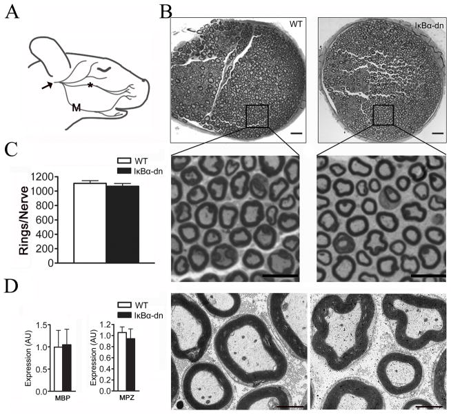
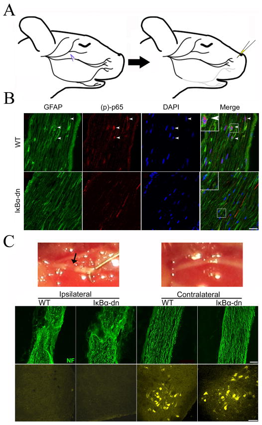
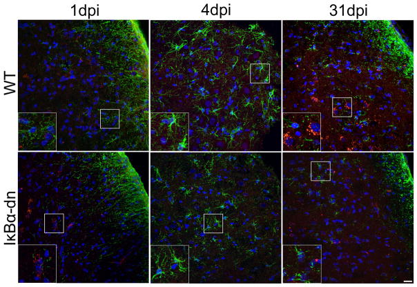
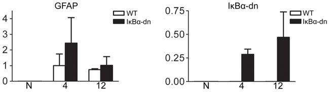
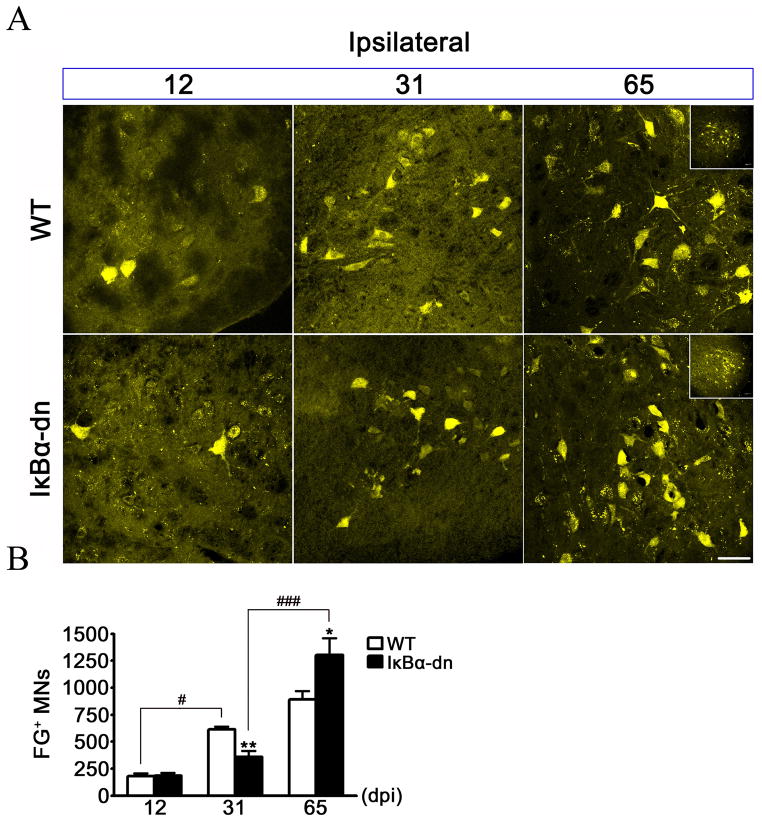
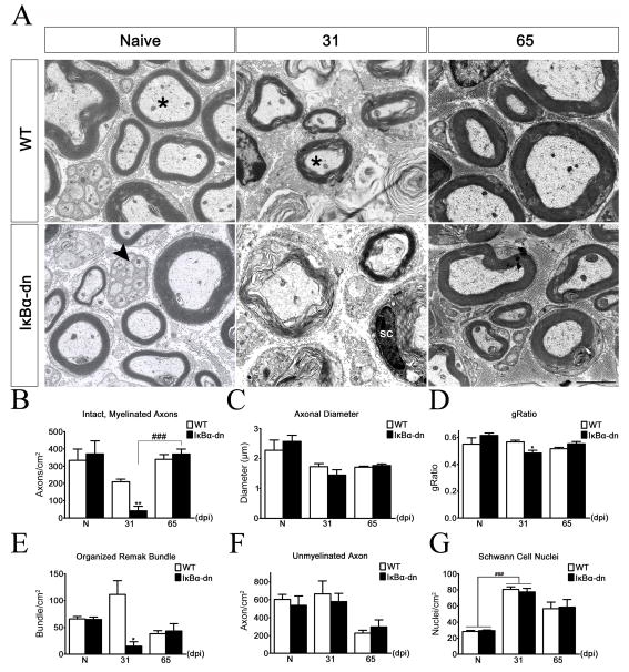
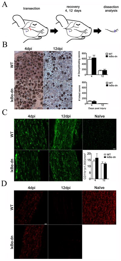
Schwann cells (SCs) are crucial for peripheral nerve development and regeneration; however, the intrinsic regulatory mechanisms governing postinjury responses are poorly understood. Activation and deacetylation of nuclear factor-κB (NF- κB) in SCs have been implicated as prerequisites for peripheral nerve myelination. Using GFAP-IκBα-dn mice in which NF- κB transcriptional activation is inhibited in SCs, we found no discernable differences in the quantity or structure of myelinated axons in adult facial nerves. Following crush injury, axonal regeneration was impaired at 31 days and significantly enhanced at 65 days in transgenic animals. Compact remyelination and Remak bundle organization were significantly compromised at 31 days and restored by 65 days post injury. Together, these data indicate that inhibition of NF-κB activation in SCs transiently delays axonal regeneration and compact remyelination. Manipulating the temporal activation of nuclear factor-κB in Schwann cells may offer new therapeutic avenues for PNS and CNS regeneration.
Copyright © 2012 Wiley Periodicals, Inc.
Figures


Similar articles
-
Activation of NF-κB in Schwann cells is dispensable for myelination in vivo.J Neurosci. 2013 Jun 12;33(24):9932-6. doi: 10.1523/JNEUROSCI.2483-12.2013. J Neurosci. 2013. PMID: 23761888 Free PMC article.
-
Axonal neuregulin 1 is a rate limiting but not essential factor for nerve remyelination.Brain. 2013 Jul;136(Pt 7):2279-97. doi: 10.1093/brain/awt148. Brain. 2013. PMID: 23801741 Free PMC article.
-
A role for Schwann cell-derived neuregulin-1 in remyelination.Nat Neurosci. 2013 Jan;16(1):48-54. doi: 10.1038/nn.3281. Epub 2012 Dec 9. Nat Neurosci. 2013. PMID: 23222914
-
Models and methods to study Schwann cells.J Anat. 2022 Nov;241(5):1235-1258. doi: 10.1111/joa.13606. Epub 2022 Jan 5. J Anat. 2022. PMID: 34988978 Free PMC article. Review.
-
Wallerian degeneration: gaining perspective on inflammatory events after peripheral nerve injury.J Neuroinflammation. 2011 Aug 30;8:110. doi: 10.1186/1742-2094-8-110. J Neuroinflammation. 2011. PMID: 21878126 Free PMC article. Review.
Cited by
-
Nerve injury induces glial cell line-derived neurotrophic factor (GDNF) expression in Schwann cells through purinergic signaling and the PKC-PKD pathway.Glia. 2013 Jul;61(7):1029-40. doi: 10.1002/glia.22491. Epub 2013 Apr 2. Glia. 2013. PMID: 23553603 Free PMC article.
-
Activation of NF-κB in Schwann cells is dispensable for myelination in vivo.J Neurosci. 2013 Jun 12;33(24):9932-6. doi: 10.1523/JNEUROSCI.2483-12.2013. J Neurosci. 2013. PMID: 23761888 Free PMC article.
-
Inhibition of NADPH oxidase activation in oligodendrocytes reduces cytotoxicity following trauma.PLoS One. 2013 Nov 19;8(11):e80975. doi: 10.1371/journal.pone.0080975. eCollection 2013. PLoS One. 2013. PMID: 24260524 Free PMC article.
-
Heparin-based coacervate of bFGF facilitates peripheral nerve regeneration by inhibiting endoplasmic reticulum stress following sciatic nerve injury.Oncotarget. 2017 Jul 18;8(29):48086-48097. doi: 10.18632/oncotarget.18256. Oncotarget. 2017. PMID: 28624802 Free PMC article.
-
Negative regulators of schwann cell differentiation-novel targets for peripheral nerve therapies?J Clin Immunol. 2013 Jan;33 Suppl 1:S18-26. doi: 10.1007/s10875-012-9786-9. Epub 2012 Sep 6. J Clin Immunol. 2013. PMID: 22956147 Review.
References
-
- Beg AA, Sha WC, Bronson RT, Ghosh S, Baltimore D. Embryonic lethality and liver degeneration in mice lacking the RelA component of NF-kappa B. Nature. 1995;376:167–170. - PubMed
-
- Boyle K, Azari MF, Cheema SS, Petratos S. TNFalpha mediates Schwann cell death by upregulating p75NTR expression without sustained activation of NFkappaB. Neurobiol Dis. 2005;20(2):412–27. - PubMed
-
- Bracchi-Ricard V, Brambilla R, Levenson J, Hu WH, Bramwell A, Sweatt JD, Green EJ, Bethea JR. Astroglial nuclear factor-kappaB regulates learning and memory and synaptic plasticity in female mice. J Neurochem. 2008;104(3):611–23. - PubMed
Publication types
MeSH terms
Substances
Grants and funding
LinkOut - more resources
Full Text Sources
Molecular Biology Databases
Miscellaneous
