. 2012 Dec;72(12):1498-515.
doi: 10.1002/dneu.22011. Epub 2012 Jul 27.

C. elegans dystroglycan coordinates responsiveness of follower axons to dorsal/ventral and anterior/posterior guidance cues

Affiliations

C. elegans dystroglycan coordinates responsiveness of follower axons to dorsal/ventral and anterior/posterior guidance cues

Robert P Johnson et al. Dev Neurobiol. 2012 Dec.

Abstract

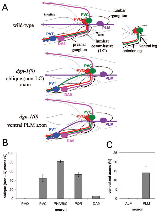
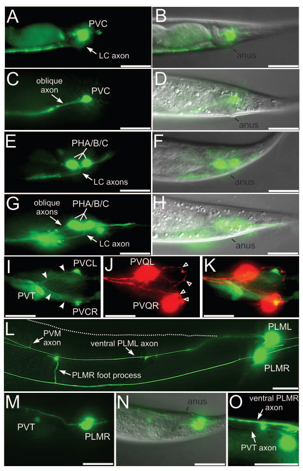
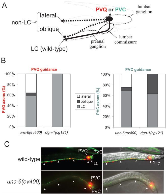
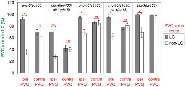
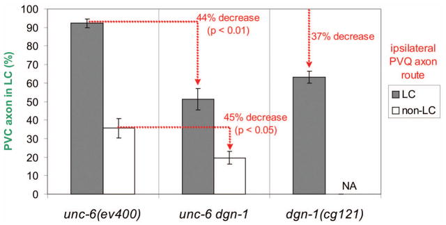
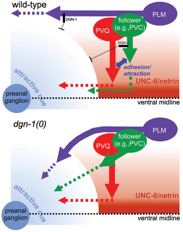
Neural development in metazoans is characterized by the establishment of initial process tracts by pioneer axons and the subsequent extension of follower axons along these pioneer processes. Mechanisms governing the fidelity of follower extension along pioneered routes are largely unknown. In C. elegans, formation of the right angle-shaped lumbar commissure connecting the lumbar and preanal ganglia is an example of pioneer/follower dynamics. We find that the dystroglycan ortholog DGN-1 mediates the fidelity of follower lumbar commissure axon extension along the pioneer axon route. In dgn-1 mutants, the axon of the pioneer PVQ neuron faithfully establishes the lumbar commissure, but axons of follower lumbar neurons, such as PVC, frequently bypass the lumbar commissure and extend along an oblique trajectory directly toward the preanal ganglion. In contrast, disruption of the UNC-6/netrin guidance pathway principally perturbs PVQ ventral guidance to pioneer the lumbar commissure. Loss of DGN-1 in unc-6 mutants has a quantitatively similar effect on follower axon guidance regardless of PVQ axon route, indicating that DGN-1 does not mediate follower/pioneer adhesion. Instead, DGN-1 appears to block premature responsiveness of follower axons to a preanal ganglion-directed guidance cue, which mediates ventral-to-anterior reorientation of lumbar commissure axons. Deletion analysis shows that only the most N-terminal DGN-1 domain is required for these activities. These studies suggest that dystroglycan modulation of growth cone responsiveness to conflicting guidance cues is important for restricting follower axon extension to the tracts laid down by pioneers.


Figures

Figure 1
Figure 1. Defects in lumbar ganglion neural guidance in dgn-1 mutants
(A) Several lumbar ganglion neurons, including PVQ (red) and PVC (green), project axons into the preanal ganglion through the lumbar commissure (LC), which has distinct ventral and anterior legs. The PLM (purple) axon in contrast projects anteriorly at a lateral position. The preanal ganglion neurons DA8 (not shown) and DA9 (pink) project axons dorsally through the left and right lumbar commissures, respectively; for convenience, DA9 is shown on the left in this schematic (upper panel). In dgn-1 null mutants, follower LC axons such as PVC sometimes fail to enter the lumbar commissure, instead taking an oblique route toward the preanal ganglion (middle panel), and PLM axons are occasionally directed ventrally toward the preanal ganglion (lower panel). (B) Oblique (non-LC) defects of specific lumbar commissure axons in dgn-1(cg121) null mutants is shown. For PHA/B/C, one or more of three axons per side could be defective (see Materials and Methods). (C) Ventralization of PLM axons in dgn-1(cg121). In B and C, percent of axons with guidance defects and the standard error of the proportion (error bars) is shown; N=100–232 axons scored for each neuron type.
Figure 2
Figure 2. Axon guidance defects in lumbar neurons of dgn-1 mutants
(A–D) Lateral views of lumbar commissure PVC (A) or PHA/B/C (E) axons in wild-type animals, and of oblique PVC (C) or PHA/B/C (G) axon trajectories in dgn-1(cg121) animals. One of the three PHA/B/C axons extends normally through the lumbar commissure in G. Overlays with the corresponding DIC images in (B,D,F,H) marking position of the anus. (I–K) Ventral view of a dgn-1(cg121) animal carrying markers for PVC (green, I), PVQ (red, J) and the preanal ganglion neuron PVT (green, I). Oblique PVC axons (I, filled arrowheads) extend directly toward the preanal ganglion, marked by PVT. The pioneer PVQ axons always traverse the lumbar commissure (J, open arrowheads) in dgn-1 mutants. K, overlay of I and J. In dgn-1(cg121), lumbar neuron cell bodies can be displaced anterior of their normal position, e.g. PVQL in J. (L) PLML axon in dgn-1(cg121) misrouted into the ventral nerve cord (marked by the entering PVM axon and the ventral cord foot process of PLMR). Normal lateral course of PLML is indicated (dotted line). In contrast the PLMR axon follows the normal lateral trajectory. (M–O) Subventral view of a dgn-1(cg121) animal carrying markers for PLM and PVT (green, M) and overlay with the DIC image (N), showing the misguided PLMR axon entering the ventral cord in the preanal ganglion region between PVT and the anus. In a different focal plane (O), the PVT and PLMR axons can be seen running parallel in the ventral cord. Bars: 20 μm.
Figure 3
Figure 3. Embryonic expression of dgn-1 in lumbar ganglion neurons
(A) In comma stage embryos, a dgn-1p::GFP transcriptional reporter is expressed in several lumbar ganglion (lg) neurons. (B) Overlay of A with corresponding differential interference contrast (DIC) image. (C,D) Enlargement of boxed regions in A,B. The lumbar ganglion neurons are the most posterior neurons (identified by a stippled appearance under DIC) in the embryo. (E) Expression of dgn-1p::GFP can still be observed in early L1 stage animals in some lumbar neurons (PVQ, PLM, ALN) as well as in the rectal gland cells (rect). (F) DIC image of E. Bar: 20 μm (A,B,E,F) or 10 μm (C,D).
Figure 4
Figure 4. Rescue of lumbar commissure guidance in dgn-1 mutants by early neural expression of dgn-1(+)
PVC oblique guidance defects were scored in dgn-1(cg121) or wild-type animals carrying an extrachromosomal array containing dgn-1(+) transgenes driven by the indicated heterologous promoters. Average and range (error bars) of two independent extrachromosomal array lines is shown. N=100–138 axons scored for each transgene line in each genetic background. Some averages or ranges are 0 and no bar is visible.
Figure 5
Figure 5. Function of DGN-1 structural domains in axon guidance and gonadogenesis
A series of DGN-1 domain deletion mutants (A, left panel) was expressed transgenically in dgn-1(cg121) or wild-type animals under the control of the early neural promoter unc-119p. Transgenic animals were scored for oblique PVC axons (A, right panel), ventralized PLM axons (B), and sterility (C). Average and range (error bars) of two independent extrachromosomal array lines is indicated. N=100–150 axons (A,B) or N=50–75 animals (C) scored for each transgene line in each genetic background.
Figure 6
Figure 6. Distinct lumbar guidance defects in unc-6/netrin and dgn-1/dystroglycan mutants
(A) PVQ and PVC axons were scored for normal transit of the lumbar commissure (LC) or failure to transit the lumbar commissure by either following an oblique trajectory toward the preanal ganglion or by extending at a lateral position and failing to enter the preanal ganglion. (B) Distribution of PVQ and PVC axon trajectories in unc-6/netrin (N=200 axons for each neuron) and dgn-1/dystroglycan mutants (N=212 axons for each neuron). (C) Example of lateralized PVQ and PVC axons in unc-6/netrin mutants. In wild-type animals PVQ (red) and PVC (green) axons extend ventrally through the lumbar commissure (LC) and anteriorly along the ventral nerve cord at the ventral surface (arrowheads). In unc-6(ev400) netrin mutants, ventral guidance of PVQ and PVC axons sometimes fails completely, and axons extend directly anterior at a lateral position, away from the ventral surface (arrowheads).
Figure 7
Figure 7. Ipsilateral PVQ pioneer axon route is the major determinant of follower axon trajectory
PVC axon route (LC or non-LC as in Fig. 6A) in netrin and Slit pathway mutants was correlated with the route taken by either the ipsilateral (ipsi PVQ) or the contralateral (contra PVQ) PVQ axon. Data analyzed by a two-tailed Z test: *, p < 0.002; ns, not significant. N=76–215 ipsilateral or contralateral PVC/PVQ pairs scored for either PVQ route (LC or non-LC) in each mutant background, except for sax-3(ky123), where N=1422 (LC PVQ) or N=26 (non-LC PVQ) ipsilateral or contralateral PVC/PVQ pairs. PVC vs. contralateral PVQ data for sax-3(ky123) could not be analyzed for significance because the small absolute number of non-LC PVC observed (2/26) fell below the recommended threshold for Z test application.
Figure 8
Figure 8. DGN-1 is required for both PVQ-dependent and PVQ-independent guidance of follower lumbar axons
The fidelity of lumbar commissure guidance of PVC follower axons was correlated with the route (LC or non-LC) of the ipsilateral PVQ axon in unc-6(ev400), dgn-1(cg121) and unc-6 dgn-1 double mutants. Loss of dgn-1 in the unc-6 background reduces the fidelity of PVC guidance to a similar extent regardless of ipsilateral PVQ route. Average and standard error of the proportion (error bars) is shown. N= 78–212 ipsilateral PVC/PVQ pairs scored for either PVQ route (LC or non-LC) in each mutant background. NA, not applicable: dgn-1(cg121) shows no non-LC trajectories for PVQ.
Figure 9
Figure 9. DGN-1/dystroglycan suppresses premature or abnormal response of lumbar axons to a local guidance cue
In wild-type animals (upper panel), UNC-6/netrin signaling directs PVQ axon extension ventrally to the midline, where a local guidance cue, probably produced by the preanal ganglion, promotes anterior turning of the PVQ growth cone. In follower lumbar neurons, DGN-1 acts to block premature response to the preanal ganglion cue, allowing the follower axons to track along the PVQ pioneer through an adhesive or short-range attractive interaction. In the absence of DGN-1 (lower panel), follower lumbar axons polarize obliquely toward the preanal ganglion cue rather than tracking along PVQ. DGN-1 similarly blocks abnormal response of the PLM axon to the same attractive preanal ganglion signal, allowing it to maintain a lateral position.

Similar articles

Cited by

References

    1. Akhavan A, Crivelli SN, Singh M, Lingappa VR, Muschler JL. SEA domain proteolysis determines the functional composition of dystroglycan. FASEB J. 2008;22:612–21. - PubMed
    1. Altun-Gultekin Z, Andachi Y, Tsalik EL, Pilgrim D, Kohara Y, Hobert O. A regulatory cascade of three homeobox genes, ceh-10, ttx-3 and ceh-23, controls cell fate specification of a defined interneuron class in C. elegans. Development. 2001;128:1951–69. - PubMed
    1. Aurelio O, Hall DH, Hobert O. Immunoglobulin-domain proteins required for maintenance of ventral nerve cord organization. Science. 2002;295:686–90. - PubMed
    1. Bak M, Fraser SE. Axon fasciculation and differences in midline kinetics between pioneer and follower axons within commissural fascicles. Development. 2003;130:4999–5008. - PubMed
    1. Benard C, Tjoe N, Boulin T, Recio J, Hobert O. The small, secreted immunoglobulin protein ZIG-3 maintains axon position in Caenorhabditis elegans. Genetics. 2009;183:917–27. - PMC - PubMed

Publication types

MeSH terms

Substances