

Neogenin regulates Sonic Hedgehog pathway activity during digit patterning
- PMID: 22275192
- PMCID: PMC3424067
- DOI: 10.1002/dvdy.23745
Neogenin regulates Sonic Hedgehog pathway activity during digit patterning
Abstract
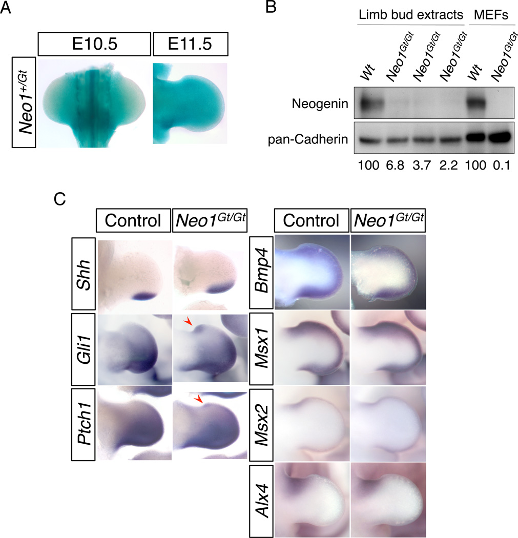
Background: Digit patterning integrates signaling by the Sonic Hedgehog (SHH), fibroblast growth factor (FGF), and bone morphogenetic protein (BMP) pathways. GLI3, a component of the SHH pathway, is a major regulator of digit number and identity. Neogenin (encoded by Neo1) is a cell surface protein that serves to transduce signals from several ligands, including BMPs, in various developmental contexts. Although neogenin is implicated in BMP signaling, it has not been linked to SHH signaling and its role in digit patterning is unknown.
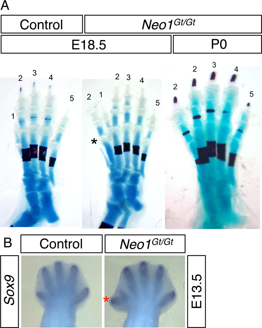
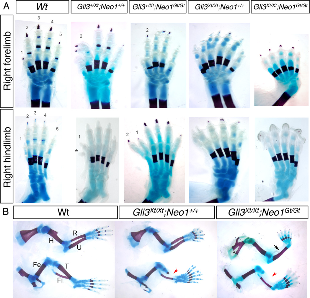
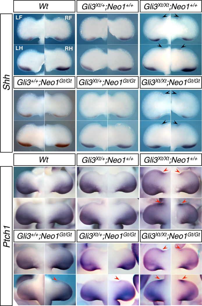
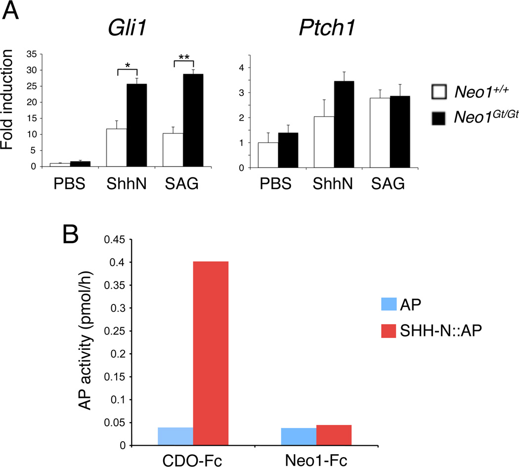
Results: We report that Neo1 mutant mice have preaxial polydactyly with low penetrance. Expression of SHH target genes, but not BMP target genes, is altered in Neo1 mutant limb buds. Analysis of mice carrying mutations in both Neo1 and Gli3 reveals that, although neogenin plays a role in constraint of digit numbers, suppressing polydactyly, it is also required for the severe polydactyly caused by loss of GLI3. Furthermore, embryo fibroblasts from Neo1 mutant mice are sensitized to SHH pathway activation in vitro.
Conclusions: Our findings indicate that neogenin regulates SHH signaling in the limb bud to achieve proper digit patterning.
Copyright © 2012 Wiley-Liss, Inc.
Figures
Similar articles
-
Preaxial polydactyly caused by Gli3 haploinsufficiency is rescued by Zic3 loss of function in mice.Hum Mol Genet. 2012 Apr 15;21(8):1888-96. doi: 10.1093/hmg/dds002. Epub 2012 Jan 10. Hum Mol Genet. 2012. PMID: 22234993 Free PMC article.
-
Suppressor of Fused Is Required for Determining Digit Number and Identity via Gli3/Fgfs/Gremlin.PLoS One. 2015 May 22;10(5):e0128006. doi: 10.1371/journal.pone.0128006. eCollection 2015. PLoS One. 2015. PMID: 26001200 Free PMC article.
-
Progression of vertebrate limb development through SHH-mediated counteraction of GLI3.Science. 2002 Oct 25;298(5594):827-30. doi: 10.1126/science.1075620. Epub 2002 Sep 5. Science. 2002. PMID: 12215652
-
Developmental biology of the upper limb.Hand Surg Rehabil. 2018 Oct;37(5):265-274. doi: 10.1016/j.hansur.2018.03.007. Epub 2018 Jul 21. Hand Surg Rehabil. 2018. PMID: 30041930 Review.
-
Control of murine kidney development by sonic hedgehog and its GLI effectors.Cell Cycle. 2006 Jul;5(13):1426-30. doi: 10.4161/cc.5.13.2928. Epub 2006 Jul 1. Cell Cycle. 2006. PMID: 16855389 Review.
Cited by
-
Transcriptomic profile adaptations following exposure of equine satellite cells to nutriactive phytochemical gamma-oryzanol.Genes Nutr. 2016 Mar 17;11:5. doi: 10.1186/s12263-016-0523-5. eCollection 2016. Genes Nutr. 2016. PMID: 27482297 Free PMC article.
-
Neogenin in Amygdala for Neuronal Activity and Information Processing.J Neurosci. 2018 Oct 31;38(44):9600-9613. doi: 10.1523/JNEUROSCI.0433-18.2018. Epub 2018 Sep 18. J Neurosci. 2018. PMID: 30228230 Free PMC article.
-
The Polygenic Map of Keloid Fibroblasts Reveals Fibrosis-Associated Gene Alterations in Inflammation and Immune Responses.Front Immunol. 2022 Jan 10;12:810290. doi: 10.3389/fimmu.2021.810290. eCollection 2021. Front Immunol. 2022. PMID: 35082796 Free PMC article.
-
Neogenin-loss in neural crest cells results in persistent hyperplastic primary vitreous formation.J Mol Cell Biol. 2020 Jan 22;12(1):17-31. doi: 10.1093/jmcb/mjz076. J Mol Cell Biol. 2020. PMID: 31336386 Free PMC article.
-
Hippocampal astrocytic neogenin regulating glutamate uptake, a critical pathway for preventing epileptic response.Proc Natl Acad Sci U S A. 2021 Apr 20;118(16):e2022921118. doi: 10.1073/pnas.2022921118. Proc Natl Acad Sci U S A. 2021. PMID: 33850017 Free PMC article.
References
-
- De Vries M, Cooper HM. Emerging roles for neogenin and its ligands in CNS development. J Neurochem. 2008;106:1483–1492. - PubMed
Publication types
MeSH terms
Substances
Grants and funding
LinkOut - more resources
Full Text Sources
Molecular Biology Databases
