. 2012 Feb;241(2):376-89.
doi: 10.1002/dvdy.23717. Epub 2012 Jan 3.

Notch pathway regulation of neural crest cell development in vivo

Affiliations

Notch pathway regulation of neural crest cell development in vivo

Timothy J Mead et al. Dev Dyn. 2012 Feb.

Abstract

Background: The function of Notch signaling in murine neural crest-derived cell lineages in vivo was examined.

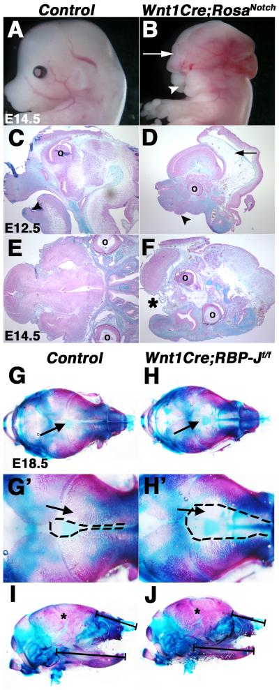
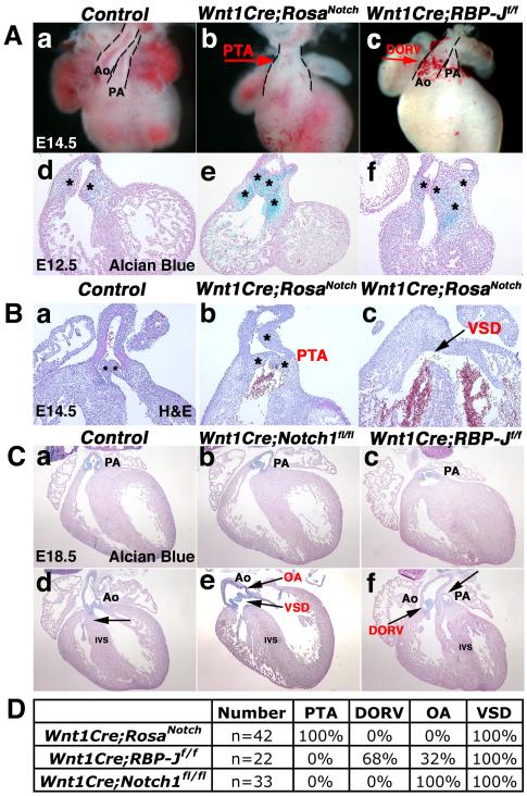
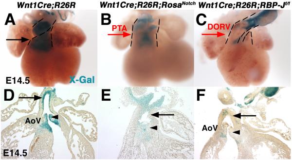
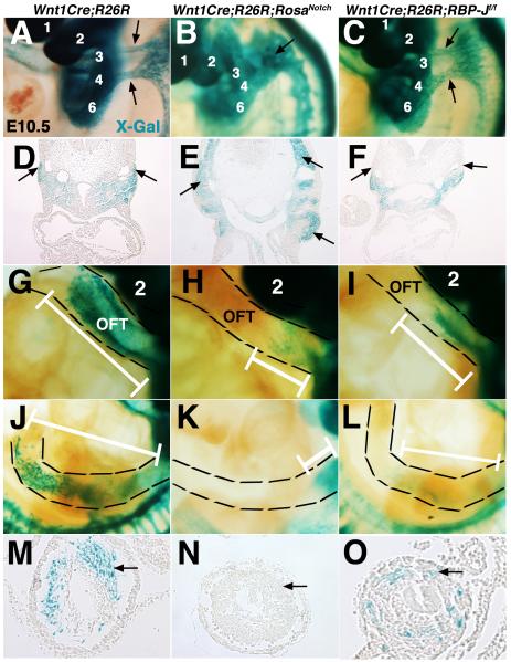
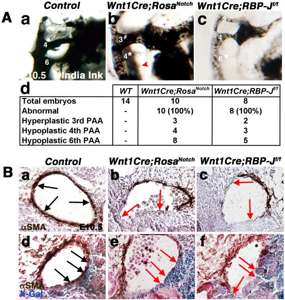
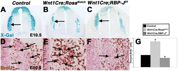
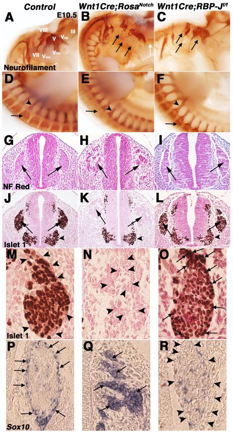
Results: Conditional gain (Wnt1Cre;Rosa(Notch)) or loss (Wnt1Cre;RBP-J(f/f)) of Notch signaling in neural crest cells (NCCs) in vivo results in craniofacial, cardiac, and trunk abnormalities. Severe craniofacial malformations are apparent in Wnt1Cre;Rosa(Notch) embryos, while less severe skull abnormalities are evident in Wnt1Cre;RBP-J(f/f) mice. Deficient cardiac neural crest migration, resulting in cardiac outflow tract malformations, occurs with increased or decreased Notch signaling in NCCs. Smooth muscle cell differentiation also is impaired in pharyngeal NCC derivatives in both Wnt1Cre;Rosa(Notch) and Wnt1Cre;RBP-J(f/f) embryos. Neurogenesis is absent and gliogenesis is increased in the dorsal root ganglia of Wnt1Cre;Rosa(Notch) embryos, while neurogenesis is increased and gliogenesis is decreased in Wnt1Cre;RBP-J(f/f) embryos.

Conclusions: Together, these studies demonstrate essential cell-autonomous roles for appropriate levels of Notch signaling during NCC migration, proliferation, and differentiation with critical implications in craniofacial, cardiac, and neurogenic development and disease.


Figures

Fig. 1
Fig. 1
Craniofacial malformations result from activation or inactivation of Notch signaling in the neural crest. (A-F) Wnt1Cre;RosaNotch embryos (n=4) exhibit craniofacial malformations, consisting of exencephaly (B, D, arrows), micrognathia (D, arrowhead), cleft face (B, arrowhead) and palate (F, asterisk). Embryos are shown in whole mount (A,B) and in Alcian blue and nuclear fast red counter stained cranial sections (C-F). (G-H’) Alcian blue and Alizarin red stained cranial skeletal preparations of E18.5 Wnt1Cre;RBP-Jf/f embryos exhibit expanded frontal cranial sutures as a result of deficient formation of craniofacial frontal bones apparent in reduced Alizarin red-stained structures, as compared to a wildtype control littermate (arrows) (n=4). (I-J) Mandible size and nasal cartilage are reduced in Wnt1Cre;RBP-Jf/f embryos (n=6), as compared to a wildtype control littermate (n=6). ° denotes eye placement. Dotted lines (G’, H’) represent the area of proximal frontal bones. * indicate apparently normal coronal suture closure (I,J). Black brackets (I, J) denote length of mandible and nasal cartilage.
Fig. 2
Fig. 2
Neural crest-specific gain or loss of Notch signaling results in cardiac outflow tract malformations. (A) Wnt1Cre;RosaNotch hearts exhibit defective cardiac OFT septation resulting in PTA (b, red arrow; e). DORV is apparent in Wnt1Cre;RBP-Jf/f hearts (c, red arrow; f) as compared to a wildtype control littermate (a,d). Endocardial cushions are stained with Alcian blue (d-f). (B) Wnt1Cre;RosaNotch hearts exhibit PTA and an accompanied VSD (black arrow) at E14.5 apparent in H&E stained sections as compared to a Wnt1Cre-negative RosaNotch control littermate (a-c). (C) Wnt1Cre;Notch1fl/fl hearts exhibit OA and VSD (e, black arrows), while Wnt1Cre;RBP-Jf/f heart sections exhibit DORV with pulmonary stenosis (f, black arrows), as compared to E18.5 control sections (a,d). The intact IVS is indicated by an arrow in d. Valve leaflets are stained with Alcian blue (a-f). (D) Wnt1Cre;RosaNotch mice exhibit 100% penetrance of PTA and VSD at E14.5. 100% of Wnt1Cre;RBP-Jf/f embryos have OFT malformations with the majority containing DORV and the minority exhibiting OA from E14.5 until birth. Wnt1Cre;Notch1fl/fl mice exhibit 100% OA from E14.5 until birth. Dashed lines outline OFT (A). * Denotes number of heart cushions in section plane (B). Ao, aortic root; PA, pulmonary artery root; PTA, persistent truncus arteriosus; DORV, double outlet right ventricle; VSD, ventricular septal defect; OA, overriding aorta; IVS, interventricular septum. (n=8 for each genotype at each age analyzed)
Fig. 3
Fig. 3
The presence of cardiac neural crest cell derivatives is reduced in the developing OFT in mice with gain or loss of Notch signaling in the NCC associated with congenital cardiac malformations. Expression of β-gal from the R26R allele was detected by blue X-gal staining of isolated hearts. Wnt1Cre;R26R;RosaNotch (B,E) and Wnt1Cre;R26R;RBP-Jf/f (C,F) embryos have reduced presence of neural crest-derived cells in the cardiac outflow tract and aortic valves at E14.5, as compared to Wnt1Cre;R26R controls (A, D, arrows, arrowheads). Dashed lines outline the outflow tract. PTA, persistent truncus arteriosus; DORV, double outlet right ventricle; AoV, aortic valve. (n=6 for each genotype at each age analyzed)
Fig. 4
Fig. 4
Cardiac and trunk neural crest cell migration is defective in mice with gain or loss of Notch signaling in neural crest derivatives. (A-F) X-Gal-stained E10.5 embryos and sections illustrate abnormal NCC migration into PA3 and 4 of Wnt1Cre;R26R;RosaNotch embryos as compared to two streams of migrating cardiac NCCs in the Wnt1Cre;R26R control (A, B, D, E, arrows). (G-I) Reduced cardiac NCC contribution is apparent in the Wnt1Cre;RosaNotch and Wnt1Cre;RBP-Jf/f OFT, as compared to Cre-negative control littermates. (J-O) Reduction of NCC contribution in the enteric nervous system of the gut is apparent in Wnt1Cre;RosaNotch and Wnt1Cre;RBP-Jf/f embryos (arrows). OFT, outflow tract. White brackets show migration distance. Dashed lines outline the OFT (G-I) and enteric nervous system of the midgut (J-L). PAs are labeled numerically in panels A-C and G-I. (n=6 for each genotype at each age analyzed)
Fig. 5
Fig. 5
Pharyngeal arch artery malformations and deficient smooth muscle cell differentiation are evident in E10.5 Wnt1Cre;RosaNotch and Wnt1Cre;RBP-Jf/f embryos. (A) Ink injected intracardially into E10.5 embryos illustrates pharyngeal arch artery (PAA) defects including hyperplastic 3rd PAA, hypoplastic 4th PAA, and hypoplasia of the 6th PAA (red arrowhead) in Wnt1Cre;RosaNotch embryos (b). Hypoplasia of the 6th PAA is apparent in Wnt1Cre;RBP-Jf/f hearts (c), as compared to Cre-negative control littermates (a). The frequency and spectrum of PAA defects in Wnt1Cre;RosaNotch and Wnt1Cre;RBP-Jf/f embryos with number of embryos analyzed is shown in (d). All of the mutant embryos were affected with one or more PAA abnormality and the incidence of specific PAA malformations is indicated. (B) Decreased PAA4 anti-αSMA reactivity is apparent in Wnt1Cre;RosaNotch and Wnt1Cre;RBP-Jf/f embryos, as compared to controls (a-c, arrows). Double labeling of X-Gal labeled NCCs and anti-αSMA illustrates a lack of NC-derived smooth muscle cell differentiation in PAA3, as compared to controls (d-f, arrows). # increased PAA size; * reduced PAA size; hypo, hypoplastic. (n=6 for each genotype)
Fig. 6
Fig. 6
Notch signaling promotes cell proliferation of neural crest derivatives. (A-C) The DRG (arrows) are NCC-derived structures, as evident by X-Gal staining of Wnt1Cre;R26R, Wnt1Cre;R26R;RosaNotch, and Wnt1Cre;R26R;RBP-Jf/f E10.5 sections. (n=8 for each genotype) (D-G) Notch activation results in increased NCC proliferation, while Notch pathway inactivation results in significant reduction of NCC proliferation in the DRG, as determined by BrdU incorporation (arrows), compared to Wnt1Cre-negative control littermates. (n=6 for each genotype) * p ≤ 0.01
Fig. 7
Fig. 7
Notch signaling regulates neuronal and glial cell fates in cranial and dorsal root gangliogenesis. (A-F) Whole-mount anti-neurofilament 2H3 staining illustrates defective and reduced V, VII, and VIII cranial ganglia branching (black arrows) and loss of III formation (white arrow) in a Wnt1Cre;RosaNotch embryo (B) compared to a Wnt1Cre-negative RosaNotch control littermate (A). Wnt1Cre;RosaNotch embryos also lack dorsal root ganglia (DRG) neurofilament staining (arrow in E) at E10.5. Neurofilament patterning appears normal in Wnt1Cre;RBP-Jf/f embryos but specific neuronal projections appear to be reduced in size (C, F, arrows), as compared to wildtype littermate controls (A, D). (n=5 embryos were analyzed for each genotype) Arrowheads in D-F note spinal nerve neurofilament marker expression. (G-I) Nuclear fast red stained sections of E10.5 Wnt1Cre;RosaNotch embryos demonstrate defective DRG morphogenesis (H, arrows), while Wnt1Cre;RBP-Jf/f DRG appear normal (I, arrows). (J-L) Lack of neurogenic marker Islet1 protein expression is apparent in Wnt1Cre;RosaNotch DRG (K, arrows). Arrowheads in J-L denote Islet1 expression in non-NC derived motor neurons. (M-R) 60x objective magnification of control DRG show Islet1-positive cells in the center of the DRG with Sox10-expressing glial cells surrounding (M, P, arrowheads). Wnt1Cre;RosaNotch DRG lack Islet1 (N, arrowheads), but have increased Sox10 expression in the DRG (Q, arrows). Wnt1Cre;RBP-Jf/f DRG have reduced Sox10 (R, arrowheads) and increased Islet1 expression (O, arrows) in the DRG outer cell layer. (n=6 embryos were analyzed for each genotype)

Similar articles

Cited by

References

    1. Bergwerff M, DeRuiter MC, Hall S, Poelmann RE, Gittenberger-de Groot AC. Unique vascular morphology of the fourth aortic arches: possible implications for pathogenesis of type-B aortic arch interruption and anomalous right subclavian artery. Cardiovasc. Res. 1999;44:185–196. - PubMed
    1. Bolos V, Grego-Bessa J, de la Pompa JL. Notch signaling in development and cancer. Endocr. Rev. 2007;28:339–363. - PubMed
    1. Britsch S, Goerich DE, Riethmacher D, Peirano RI, Rossner M, Nave KA, Birchmeier C, Wegner M. The transcription factor Sox10 is a key regulator of peripheral glial development. Genes Dev. 2001;15:66–78. - PMC - PubMed
    1. Chai Y, Jiang X, Ito Y, Bringas P, Jr, Han J, Rowitch DH, Soriano P, McMahon AP, Sucov HM. Fate of the mammalian cranial neural crest during tooth and mandibular morphogenesis. Development. 2000;127:1671–1679. - PubMed
    1. Cheng HT, Kim M, Valerius MT, Surendran K, Schuster-Gossler K, Gossler A, McMahon AP, Kopan R. Notch2, but not Notch1, is required for proximal fate acquisition in the mammalian nephron. Development. 2007;134:801–811. - PMC - PubMed

Publication types

LinkOut - more resources