

Heritability of articular cartilage regeneration and its association with ear wound healing in mice
- PMID: 22275233
- PMCID: PMC3360138
- DOI: 10.1002/art.34396
Heritability of articular cartilage regeneration and its association with ear wound healing in mice
Abstract
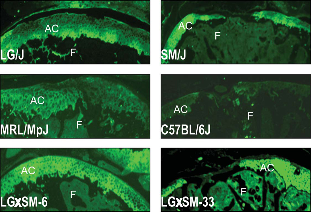
Objective: Emerging evidence suggests that genetic components contribute significantly to cartilage degeneration in osteoarthritis pathophysiology, but little information is available on the genetics of cartilage regeneration. Therefore, this study was undertaken to investigate cartilage regeneration in genetic murine models using common inbred strains and a set of recombinant inbred (RI) lines generated from LG/J (healer of ear wounds) and SM/J (nonhealer) inbred mouse strains.
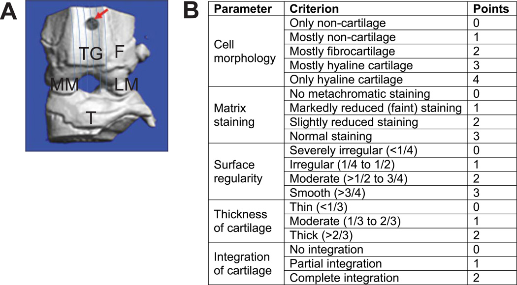
Methods: An acute full-thickness cartilage injury was introduced in the trochlear groove of 8-week-old mice (n=265) through microsurgery. Mouse knee joints were sagittally sectioned and stained with toluidine blue to evaluate regeneration. For the ear wound phenotype, a bilateral 2-mm through-and-through puncture was created in 6-week-old mice (n=229), and healing outcomes were measured after 30 days. Broad-sense heritability and genetic correlations were calculated for both phenotypes.
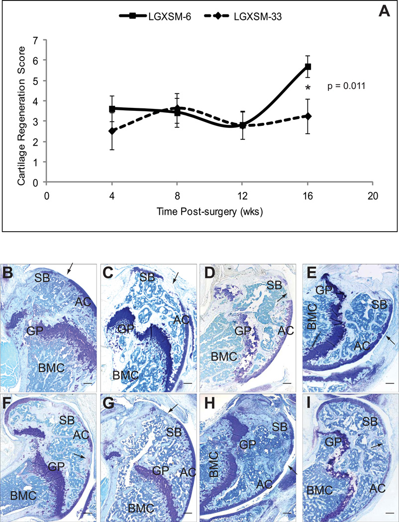
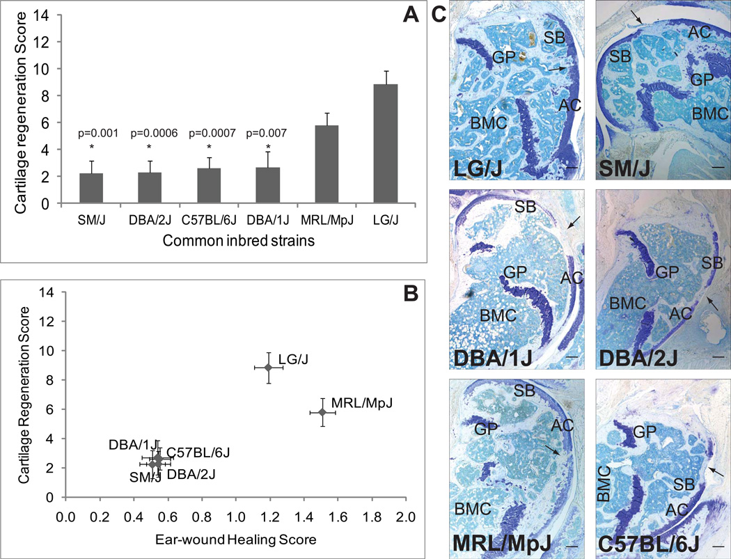
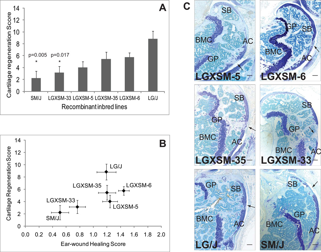
Results: Time-course analysis of the RI mouse lines showed no significant regeneration until 16 weeks after surgery; at that time, the strains could be segregated into 3 categories: good, intermediate, and poor healers. Analysis of heritability (H2) showed that both cartilage regeneration (H2=26%; P=0.006) and ear wound closure (H2=53%; P<0.00001) were significantly heritable. The genetic correlations between the two healing phenotypes for common inbred mouse strains (r=0.92) and RI mouse lines (r=0.86) were found to be extremely high.
Conclusion: Our findings indicate that articular cartilage regeneration in mice is heritable, the differences between the mouse lines are due to genetic differences, and a strong genetic correlation between the two phenotypes exists, indicating that they plausibly share a common genetic basis. We therefore surmise that LG/J by SM/J intercross mice can be used to dissect the genetic basis of variation in cartilage regeneration.
Copyright © 2012 by the American College of Rheumatology.
Figures
Similar articles
-
Molecular insight into the association between cartilage regeneration and ear wound healing in genetic mouse models: targeting new genes in regeneration.G3 (Bethesda). 2013 Nov 6;3(11):1881-91. doi: 10.1534/g3.113.007302. G3 (Bethesda). 2013. PMID: 24002865 Free PMC article.
-
Genetic correlations between cartilage regeneration and degeneration reveal an inverse relationship.Osteoarthritis Cartilage. 2020 Aug;28(8):1111-1120. doi: 10.1016/j.joca.2020.04.013. Epub 2020 May 11. Osteoarthritis Cartilage. 2020. PMID: 32437968 Free PMC article.
-
Cartilage and bone changes during development of post-traumatic osteoarthritis in selected LGXSM recombinant inbred mice.Osteoarthritis Cartilage. 2012 Jun;20(6):562-71. doi: 10.1016/j.joca.2012.01.022. Epub 2012 Feb 8. Osteoarthritis Cartilage. 2012. PMID: 22361237 Free PMC article.
-
Regeneration of articular cartilage in healer and non-healer mice.Matrix Biol. 2014 Oct;39:50-5. doi: 10.1016/j.matbio.2014.08.011. Epub 2014 Aug 28. Matrix Biol. 2014. PMID: 25173437 Free PMC article. Review.
-
Spallanzani's mouse: a model of restoration and regeneration.Curr Top Microbiol Immunol. 2004;280:165-89. doi: 10.1007/978-3-642-18846-6_5. Curr Top Microbiol Immunol. 2004. PMID: 14594211 Review.
Cited by
-
Using whole-genome sequences of the LG/J and SM/J inbred mouse strains to prioritize quantitative trait genes and nucleotides.BMC Genomics. 2015 May 28;16(1):415. doi: 10.1186/s12864-015-1592-3. BMC Genomics. 2015. PMID: 26016481 Free PMC article.
-
Genetic loci that regulate ectopic calcification in response to knee trauma in LG/J by SM/J advanced intercross mice.J Orthop Res. 2015 Oct;33(10):1412-23. doi: 10.1002/jor.22944. Epub 2015 Jun 19. J Orthop Res. 2015. PMID: 25989359 Free PMC article.
-
Molecular biomarker approaches to prevention of post-traumatic osteoarthritis.Nat Rev Rheumatol. 2024 May;20(5):272-289. doi: 10.1038/s41584-024-01102-y. Epub 2024 Apr 11. Nat Rev Rheumatol. 2024. PMID: 38605249 Review.
-
Enhanced cartilage repair in 'healer' mice-New leads in the search for better clinical options for cartilage repair.Semin Cell Dev Biol. 2017 Feb;62:78-85. doi: 10.1016/j.semcdb.2016.04.018. Epub 2016 Apr 26. Semin Cell Dev Biol. 2017. PMID: 27130635 Free PMC article. Review.
-
Molecular insight into the association between cartilage regeneration and ear wound healing in genetic mouse models: targeting new genes in regeneration.G3 (Bethesda). 2013 Nov 6;3(11):1881-91. doi: 10.1534/g3.113.007302. G3 (Bethesda). 2013. PMID: 24002865 Free PMC article.
References
-
- Gierer A, Berking S, Bode H, David CN, Flick K, Hansmann G, et al. Regeneration of hydra from reaggregated cells. Nat New Biol. 1972;239(91):98–101. - PubMed
-
- Murphy ED, Roths JB. Autoimmunity and lymphproliferation: Induction by mutant gene lpr and acceleration by a male-associated factor in strain BXSB. In: Rose NR, Bigazzi PE, Warner NL, editors. Genetic Control of Autoimmune Disease. New York: Elsevier; 1979. pp. 207–220.
-
- Stocum DL. The urodele limb regeneration blastema. Determination and organization of the morphogenetic field. Differentiation. 1984;27(1):13–28. - PubMed
-
- Tanaka EM. Regeneration: if they can do it, why can't we? Cell. 2003;113(5):559–562. - PubMed
-
- Harty M, Neff AW, King MW, Mescher AL. Regeneration or scarring: an immunologic perspective. Dev Dyn. 2003;226(2):268–279. - PubMed
Publication types
MeSH terms
Grants and funding
LinkOut - more resources
Full Text Sources
Molecular Biology Databases
