. 2012 Mar 9;287(11):8384-97.
doi: 10.1074/jbc.M112.341586. Epub 2012 Jan 24.

Insulin-like growth factor receptor-1 and nuclear factor κB are crucial survival signals that regulate caspase-3-mediated lens epithelial cell differentiation initiation

Affiliations

Insulin-like growth factor receptor-1 and nuclear factor κB are crucial survival signals that regulate caspase-3-mediated lens epithelial cell differentiation initiation

Subhasree Basu et al. J Biol Chem. .

Abstract

It is now known that the function of the caspase family of proteases is not restricted to effectors of programmed cell death. For example, there is a significant non-apoptotic role for caspase-3 in cell differentiation. Our own studies in the developing lens show that caspase-3 is activated downstream of the canonical mitochondrial death pathway to act as a molecular switch in signaling lens cell differentiation. Importantly, for this function, caspase-3 is activated at levels far below those that induce apoptosis. We now have provided evidence that regulation of caspase-3 for its role in differentiation induction is dependent on the insulin-like growth factor-1 receptor (IGF-1R) survival-signaling pathway. IGF-1R executed this regulation of caspase-3 by controlling the expression of molecules in the Bcl-2 and inhibitor of apoptosis protein (IAP) families. This effect of IGF-1R was mediated through NFκB, demonstrated here to function as a crucial downstream effector of IGF-1R. Inhibition of expression or activation of NFκB blocked expression of survival proteins in the Bcl-2 and IAP families and removed controls on the activation state of caspase-3. The high level of caspase-3 activation that resulted from inhibiting this IGF-1R/NFκB signaling pathway redirected cell fate from differentiation toward apoptosis. These results provided the first evidence that the IGF-1R/NFκB cell survival signal is a crucial regulator of the level of caspase-3 activation for its non-apoptotic function in signaling cell differentiation.


Figures

FIGURE 1.
FIGURE 1.
Inhibition of IGF-1R signaling leads to high levels of caspase-3 activation and blocks lens epithelial cell differentiation. Primary lens cell cultures at a stage just prior to differentiation initiation were exposed to the IGF-1R inhibitor PPP (1 μm) or to DMSO (control) for 24 h. A, phase contrast microscopy showed that PPP treatment altered lens cell morphology. B, immunoblot analysis with an antibody to p-IGF-1R showed that PPP effectively inhibited IGF-1R activation and had a minimal effect on total IGF-1R expression during the 24-h treatment period. Densitometric analyses of the immunoblots were plotted as the ratio of p-IGF-1R to IGF-1R. C, primary lens cell cultures that were exposed to the IGF-1R inhibitor PPP (1 μm) or to DMSO (control) for 24 h were immunostained for the cleaved (activated) caspase-3 17-kDa fragment (red) and co-stained with TO-PRO-3 for nuclei (blue) and F-actin (green) (shown here as an overlay with nuclei (blue)), and images were acquired by confocal microscopy. Images represent a single optical plane of 0.5 μm. Caspase-3 activation was quantified by creating fluorescence intensity line scans across the field of cells (panels i and ii), revealing increased levels of caspase-3 activation when IGF-1R activation was inhibited by PPP. PPP treatment also induced formation of pyknotic nuclei and reorganization of actin filaments in lens cells. D, the extent of caspase-3 activation also was determined by analyzing cl-PARP-1, a direct substrate of caspase-3. Immunoblot analysis showed that PARP-1 cleavage, plotted as a ratio to GAPDH loading control, increased significantly when IGF-1R activation was inhibited in lens epithelial cells. E, inhibition of IGF-1R activation blocked induction of lens differentiation-specific proteins CP49 and filensin, plotted here as a ratio to actin, demonstrating that deregulation of caspase-3 blocked its ability to function as a differentiation initiation signal. Results are representative of at least three studies. Error bars represent S.E. *, p < 0.05, t test.
FIGURE 2.
FIGURE 2.
IGF-1R mediates expression of pro-survival molecules and maintains caspase-3 at low levels that signal differentiation initiation but prevent apoptosis in lens epithelial cells. A, immunoblot analysis was performed to determine the effect of inhibiting IGF-1R activation (1 μm PPP, 24 h) on the expression of survival proteins in the Bcl-2 and IAP families including Bcl-2, Bcl-XL, p-Bad Ser-112 (compared with total Bad), ch-IAP-1, X-IAP, and survivin. Bcl-2, Bcl-XL, ch-IAP-1, and survivin were quantified and plotted as a ratio to actin. p-Bad Ser-112 and X-IAP expression were plotted as a ratio to total Bad and GAPDH, respectively. The expression of these survival proteins was suppressed when the IGF-1R signaling pathway was blocked. B, a live annexin V assay, an early apoptosis cell marker, was used as to examine whether inactivation of IGF-1R by PPP induced cell death by apoptosis. Confocal image analysis of annexin V staining showed the appearance of annexin V (green)-positive cells following exposure of the lens cell cultures to the IGF-1R inhibitor PPP. The fluorescence image of annexin V was overlaid with a differential interference contrast (DIC) image to show the annexin V-positive cells (green). C, lens cell cultures exposed as above to PPP were also examined for the induction of apoptosis by TUNEL assay (red), a late apoptotic marker. These cultures were co-stained with TO-PRO-3 to detect nuclei (blue) and evaluated by confocal microscopy. PPP treatment induced an increase in TUNEL-positive cells as compared with controls. D, E10 chicken lenses were treated in ex vivo culture with DMSO (i–iii) or PPP (iv–vi) for 24 h. 20-μm-thick cryosections of these lenses were examined following a TUNEL assay (red) and co-stained with TO-PRO-3 to detect nuclei (blue) at low and high magnification. The black-boxed area (top) in the model represents the equatorial zone (EQ) of the embryonic lens sections shown in the confocal images. The white-boxed areas in the low magnification images of DMSO-treated (i) and PPP-treated (iv) lenses represent the region in the EQ zone that is shown at higher magnification in panels ii/iii and v/vi, respectively. PPP treatment induced apoptosis of cells in the equatorial zone of the lens, which is the region of differentiation initiation in the developing lens. Arrows in panels i–iii and iv–vi denote the same cells. All confocal imaging within each study was performed using the same settings. Z-stacks were collected, and images represent a single optical plane of 0.5 μm. Results are representative of at least three independent studies. Error bars represent S.E. *, p < 0.05, t test. Scale bars, 20 μm.
FIGURE 3.
FIGURE 3.
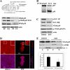
NFκB expression and activation is dependent on IGF-1R signaling in differentiating lens epithelial cells. A, E10 chicken lenses were microdissected into four distinct zones of differentiation: undifferentiated cells of the central epithelium (EC); the zone of differentiation initiation (equatorial epithelium (EQ)); cortical fiber cells (FP), where lens cell morphogenetic differentiation occurs; and nuclear fiber cells (FC), the region of fiber cell maturation. Each fraction was analyzed by immunoblot (lower panel) for expression of NFκB subunits p65 and p50 and for the activation state of NFκB p65 (p-NFκB p65 Ser-276). Samples were immunoblotted for actin as a control. A high level of expression of NFκB subunits p65 and p50 and NFκB activation occurred in the lens epithelium, with the highest in the equatorial zone of the lens. B, co-immunoprecipitation (IP) studies (immunoprecipitate for the NFκB p65 subunit and immunoblot for the NFκB p50 subunit) showed that these subunits formed a heterodimeric NFκB p50/p65 complex in the equatorial zone. WCL, whole cell lysate. C, prior to differentiation initiation, primary lens cell cultures were exposed to the IGF-1R inhibitor PPP (1 μm) or DMSO (control) for 24 h, and protein extracts were immunoblotted for active p-IGF-1R, total IGF-1R, NFκB, transcriptionally active NFκB (p-NFκB p65 Ser-276), and actin. Blocking IGF-1R activation inhibited NFκB expression and its transcriptional activity. D, the effect of exposure of lens cell cultures to PPP on translocation of the transcription factor NFκB to the nucleus was examined in lens cell cultures by confocal microscopy imaging following immunostaining for the NFκB p65 subunit (red) and co-staining with TO-PRO-3 to detect nuclei (blue). White-boxed areas show the area magnified in the inset. Images shown are of a single 0.5-μm optical plane. Inhibition of IGF-1R activation blocked the nuclear translocation of the NFκB p65 subunit. E, similarly, inhibition of the nuclear translocation of NFκB p65 was observed when IGF-1R signaling was blocked by immunoblot analysis following cell fractionation into nuclear (N) and cytoplasmic (C) cell compartments. Effectiveness of the cell nuclear/cytoplasmic fractionation was demonstrated by immunoblotting for the cytoplasmic marker GAPDH and the nuclear marker Oct-1. Densitometric analyses were performed on the level of nuclear NFκB p65 relative to Oct-1 in PPP and DMSO-treated cultures. Error bars represent S.E. *, p < 0.05, t test. Results are representative of at least three studies. Scale bar, 10 μm.
FIGURE 4.
FIGURE 4.
Blocking nuclear translocation of NFκB with the SN50 inhibitor deregulates caspase-3 activity, resulting in blocking lens epithelial cells differentiation, and down-regulates expression of survival proteins to cause apoptosis. A and B, primary lens epithelial cell cultures were exposed to the NFκB p50 inhibitor SN50 (18 μm) or DMSO (control) for 24 h, immunostained for the NFκB p50 (A) or p65 (B) subunits (red), co-stained with TO-PRO-3 to detect nuclei (blue), and examined by confocal microscopy. The white-boxed areas are shown at higher magnification in the insets. Treatment with SN50 blocked nuclear translocation of both NFκB p50 and p65 subunits. C, immunoblot analysis demonstrated that exposure to SN50 also reduced NFκB p65 expression, consistent with the self-regulation of p65. D, SN50-treated lens cells were immunostained for the cleaved (activated) caspase-3 17kD fragment (red), co-stained with TO-PRO-3 to detect nuclei (blue), and examined by confocal microscopy. The fluorescence intensity for cleaved caspase-3 (cl-caspase-3 (red)), determined by line scans created with LSM Image Examiner (i and ii) showed that exposure to SN50 increased levels of caspase-3 activation. E, this result was confirmed by immunoblot analysis for cl-PARP-1, a direct substrate for caspase-3. Immunoblot analysis also was performed to determine the effect of inhibiting NFκB nuclear translocation on the expression of lens differentiation-specific markers CP49 and filensin. F, results show that SN50 exposure inhibited lens cell differentiation. G, in addition, expression of survival proteins in the Bcl-2 and IAP families, including Bcl-2, Bcl-XL, p-Bad Ser-112, X-IAP, ch-IAP-1, and survivin, was also inhibited in the SN50-treated cells. H and I, cultures were analyzed for the effect of blocking NFκB p50 activation on lens cell survival using both annexin V (H, green) and TUNEL (I, red) assays. Nuclei in the TUNEL assay were stained with TO-PRO-3 (I, blue), and both assays were imaged by confocal microscopy. For each imaging study, analysis was performed using the same settings. Z-stacks were collected and analyzed, and the data are presented as a single 0.5-μm optical plane. Bcl-2, Bcl-XL, survivin, and X-IAP were plotted as a ratio of protein expression relative to actin; p-Bad Ser-112 and cl-PARP-1 were plotted as a ratio to total Bad and GAPDH, respectively. Error bars represent S.E. *, p < 0.05, t test. Results are representative of a minimum of three independent experiments. Scale bars, 20 μm.
FIGURE 5.
FIGURE 5.
siRNA knockdown of NFκB p65 inhibits lens epithelial cell differentiation, blocks expression of survival proteins, and induces cell death by apoptosis. A and B, prior to differentiation initiation in primary lens cell cultures, the expression of NFκB p65 was knocked down using a siRNA approach. Control cultures were exposed to non-targeting siRNA (siC). siRNA knockdown of NFκB p65 was 70% (A). Knockdown of p65 resulted in a block of phosphorylation of NFκBp65 on Ser-276, the active form of NFκB p65 (B). C, immunoblot analysis demonstrated that the knockdown of NFκB p65 blocked expression of lens-specific differentiation markers, CP49 and filensin. D, lens cell cultures immunostained for cl-caspase-3 (red) and co-stained with TO-PRO-3 to detect nuclei (blue) showed an increase in caspase-3 activation when NFκB p65 was knocked down in lens cell cultures. Fluorescence intensity of cl-caspase-3 was quantified by creating line scans (panels i and ii) across the field of cells using LSM Image Examiner, which confirmed that there was significant increase in caspase-3 activation in response to NFκB p65 knockdown. E, this increase in caspase-3 activation induced cl-PARP-1. F, expression of survival proteins in the Bcl-2 and IAP families including Bcl-2, ch-IAP-1, X-IAP, and survivin was inhibited in the absence of NFκB p65, and phosphorylation of Bad (p-BadS112) was blocked. G and H, evaluation of cell death by apoptosis in response to knockdown of NFκB p65 was performed using annexin V and TUNEL assays. Live annexin V staining illustrated an increase in annexin V-positive cells (green) when NFκB p65 was knocked down for 24 h (G). The TUNEL assay (red) co-stained with TO-PRO-3 to detect nuclei (blue); this is used as a marker for late apoptotic cell death and indicates a significant increase in the number of TUNEL-positive cells in the absence of NFκB p65 (H). Hence, siRNA knockdown of NFκB p65 inhibited the expression of survival proteins, signaling cell death instead of differentiation. Densitometric analyses of immunoblots for NFκB, filensin, CP49, survivin, ch-IAP, and Bcl-2 were plotted as a ratio relative to actin; NFκB p65 Ser-276, cl-PARP-1, and X-IAP as a ratio to GAPDH; and p-Bad Ser-112 against total Bad. For confocal imaging, Z-stacks were collected and analyzed, and the data are presented as a single optical plane of 0.5 μm. Results are representative of at least three studies. Error bars represent S.E. *, p < 0.05, t test. Scale bar, 20 μm.
FIGURE 6.
FIGURE 6.
Proposed model for the role of the IGF-1R/NFκB signaling pathway in regulating the caspase-3 differentiation initiation signal. In the differentiation of lens epithelial cells, low level activation of caspase-3 has an essential role in differentiation initiation. Caspase-3 activation for this differentiation pathway was shown previously to be downstream of the intrinsic canonical mitochondrial death pathway. Here, we show now that caspase-3 activation was maintained at the low levels at which it functions as a differentiation initiator through an IGF-1R signal that activates NFκB, a transcription factor required for the expression of survival proteins in the Bcl-2 (Bcl-2, p-Bad Ser-112, and Bcl-XL) and IAP (ch-IAP-1, X-IAP, and survivin) families. These survival proteins can regulate caspase-3 activity to maintain it at the low level required to signal lens differentiation initiation; in their absence caspase-3 is deregulated and instead signals apoptotic cell death. This is a novel role for the IGF-1R/NFκB signaling pathway in lens epithelial cell differentiation.

Similar articles

Cited by

References

    1. Maelfait J., Beyaert R. (2008) Non-apoptotic functions of caspase-8. Biochem. Pharmacol. 76, 1365–1373 - PubMed
    1. Schwerk C., Schulze-Osthoff K. (2003) Non-apoptotic functions of caspases in cellular proliferation and differentiation. Biochem. Pharmacol. 66, 1453–1458 - PubMed
    1. Weber G. F., Menko A. S. (2005) The canonical intrinsic mitochondrial death pathway has a non-apoptotic role in signaling lens cell differentiation. J. Biol. Chem. 280, 22135–22145 - PubMed
    1. Larsen B. D., Rampalli S., Burns L. E., Brunette S., Dilworth F. J., Megeney L. A. (2010) Caspase 3/caspase-activated DNase promote cell differentiation by inducing DNA strand breaks. Proc. Natl. Acad. Sci. U.S.A. 107, 4230–4235 - PMC - PubMed
    1. Fernando P., Kelly J. F., Balazsi K., Slack R. S., Megeney L. A. (2002) Caspase 3 activity is required for skeletal muscle differentiation. Proc. Natl. Acad. Sci. U.S.A. 99, 11025–11030 - PMC - PubMed

Publication types

MeSH terms