. 2012 Mar 16;287(12):9269-79.
doi: 10.1074/jbc.M111.316208. Epub 2012 Jan 24.

Stable incorporation of ATPase subunits into 19 S regulatory particle of human proteasome requires nucleotide binding and C-terminal tails

Affiliations

Stable incorporation of ATPase subunits into 19 S regulatory particle of human proteasome requires nucleotide binding and C-terminal tails

Seung-Hoon Lee et al. J Biol Chem. .

Abstract

The 26 S proteasome is a large multi-subunit protein complex that degrades ubiquitinated proteins in eukaryotic cells. Proteasome assembly is a complex process that involves formation of six- and seven-membered ring structures from homologous subunits. Here we report that the assembly of hexameric Rpt ring of the 19 S regulatory particle (RP) requires nucleotide binding but not ATP hydrolysis. Disruption of nucleotide binding to an Rpt subunit by mutation in the Walker A motif inhibits the assembly of the Rpt ring without affecting heterodimer formation with its partner Rpt subunit. Coexpression of the base assembly chaperones S5b and PAAF1 with mutant Rpt1 and Rpt6, respectively, relieves assembly inhibition of mutant Rpts by facilitating their interaction with adjacent Rpt dimers. The mutation in the Walker B motif which impairs ATP hydrolysis does not affect Rpt ring formation. Incorporation of a Walker B mutant Rpt subunit abrogates the ATPase activity of the 19 S RP, suggesting that failure of the mutant Rpt to undergo the conformational transition from an ATP-bound to an ADP-bound state impairs conformational changes in the other five wild-type Rpts in the Rpt ring. In addition, we demonstrate that the C-terminal tails of Rpt subunits possessing core particle (CP)-binding affinities facilitate the cellular assembly of the 19 S RP, implying that the 20 S CP may function as a template for base assembly in human cells. Taken together, these results suggest that the ATP-bound conformational state of an Rpt subunit with the exposed C-terminal tail is competent for cellular proteasome assembly.


Figures
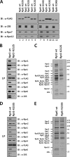
FIGURE 1.
FIGURE 1.
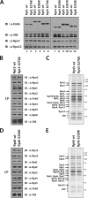
Nucleotide binding to Rpt subunits is required for the assembly of proteasome. A, impaired incorporation of Walker A mutant Rpt subunits into proteasome. HeLa cells were transiently transfected with the expression constructs of FLAG-tagged wild-type or Walker A mutant Rpt subunits as indicated. Aliquots of FLAG-Rpt samples purified in the presence of 5 mm ATP were separated by SDS-PAGE and analyzed by immunoblotting with indicated antibodies. B–C, immunoblotting (B) and SDS-PAGE (C) analyses of affinity-purified FLAG-tagged wild-type or Walker A mutant Rpt3. HeLa cell lines that conditionally expressed FLAG-tagged wild-type or Walker A mutant Rpt3 were established, and the proteasome was affinity purified as described under “Experimental Procedures.” Ten microliters each of Rpt3 samples purified in the presence of 5 mm ATP were analyzed by immunoblotting with the indicated antibodies and by SDS-PAGE followed by Coomassie staining. D and E, immunoblotting (D) and SDS-PAGE (E) analyses of affinity-purified FLAG-tagged wild-type or Walker A mutant Rpt5.
FIGURE 2.
FIGURE 2.
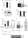
ATP hydrolysis by Rpt subunits is not essential for proteasome assembly. A, comparable incorporation of wild-type and Walker B mutant Rpt subunits into proteasome. HeLa cells were transiently transfected with the expression constructs of FLAG-tagged wild-type or Walker B mutant Rpt subunits as indicated. Aliquots of FLAG-Rpt samples purified in the presence of 5 mm ATP were separated by SDS-PAGE and analyzed by immunoblotting with the indicated antibodies. B and C, immunoblotting (B) and SDS-PAGE (C) analyses of affinity-purified FLAG-tagged wild-type or Walker B mutant Rpt3. HeLa cell lines that conditionally expressed FLAG-tagged wild-type or Walker B mutant Rpt3 were established, and the proteasome was affinity purified as described under “Experimental Procedures.” Ten microliters each of Rpt3 samples purified in the presence of 5 mm ATP was analyzed by immunoblotting with indicated antibodies and by SDS-PAGE followed by Coomassie staining. D and E, immunoblotting (D) and SDS-PAGE (E) analyses of affinity-purified FLAG-tagged wild-type or Walker B mutant Rpt6.
FIGURE 3.
FIGURE 3.
Incorporation of a single Walker B mutant Rpt subunit abrogates the ATPase activity of 19 S RP. A, comparable chymotrysin-like activities of purified proteasome samples containing the wild-type or Walker B mutant Rpt3. One microgram each of the wild-type and Walker B mutant Rpt3 samples shown in Fig. 2C was assayed for chymotrypsin-like peptidase activity using the Suc-LLVY-AMC peptide as a fluorogenic substrate. B, native PAGE analysis and substrate overlay assay of proteasome samples containing wild-type or Walker B mutant Rpt3. Affinity-purified samples were subjected to native PAGE followed by Coomassie staining or by substrate overlay assay as described under “Experimental Procedures.” C, impaired degradation of a polyubiquitinated protein substrate by proteasome containing Walker B mutant Rpt3. Polyubiquitinated T7-cIAP2-C (C2-C) was prepared as described under “Experimental Procedures.” The polyubiquitinated substrate was incubated with indicated proteasome preparations and protein degradation was analyzed by Western blotting with anti-T7 antibody. D, impaired ATPase activity of proteasome containing Walker B mutant Rpt3. FLAG-Rpt3 samples purified in the presence or absence of ATP were assayed for ATPase activity as described under “Experimental Procedures.” E, chymotrysin-like activities of proteasome samples purified with or without ATP. Wild-type and Walker B mutant FLAG-Rpt3 samples purified in the presence or absence of ATP were assayed for chymotrypsin-like peptidase activity using Suc-LLVY-AMC as a fluorogenic substrate in buffers containing the indicated concentrations of ATP. Data are presented as the mean ± S.D. from three independent experiments. F, immunoblotting and SDS-PAGE analyses of proteasome samples purified with or without ATP. Wild-type and Walker B mutant FLAG-Rpt3 samples purified in the presence or absence of ATP were analyzed by immunoblotting with indicated antibodies. SDS-PAGE analysis of proteasome samples purified in the absence of ATP is shown in the right panel. G, increased stability of 26 S proteasome during purification by AMP-PNP. Wild-type and Walker B mutant FLAG-Rpt3 samples were purified using resin binding and washing/elution buffers with or without 2 mm ATP or AMP-PNP as indicated. Samples were analyzed by immunoblotting with indicated antibodies.
FIGURE 4.
FIGURE 4.
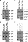
Differential effects of base-specific chaperones on proteasome assembly with Walker A mutant Rpts. A–D, immunoblotting analysis of FLAG-Rpt samples following coexpression of base-specific chaperones. HeLa cells expressing wild-type or Walker A mutant FLAG-Rpt1 (A), Rpt6 (B), Rpt3 (C), and Rpt5 (D) were transfected with the expression constructs of HA-tagged S5b, PAAF1, Gankyrin, and p27, respectively. After affinity purification of FLAG-tagged proteins in the presence of 5 mm ATP, the samples were analyzed by immunoblotting with indicated antibodies.
FIGURE 5.
FIGURE 5.
Increased interaction of an Rpt dimer containing a Walker A mutant with the adjacent dimer by S5b and PAAF1. A, immunoblotting analysis of wild-type and Walker A mutant FLAG-Rpt1 samples following expression of S5. HeLa cells were transfected with the expression constructs of wild-type or Walker A mutant FLAG-Rpt1 along with HA-tagged Rpt2, Rpt4, Rpt5, and S5B. Immunopurified FLAG-Rpt1 samples were analyzed by immunoblotting with indicated antibodies. B, immunoblotting analysis of wild-type and Walker A mutant FLAG-Rpt6 samples following the expression of PAAF1. Hela cells were transfected with the expression constructs of wild-type or Walker A mutant FLAG-Rpt6 along with HA-tagged Rpt1, Rpt2, Rpt3, and PAAF1. Immunopurified FLAG-Rpt6 samples were analyzed by immunoblotting with the indicated antibodies.
FIGURE 6.
FIGURE 6.
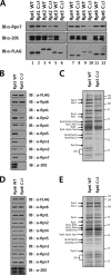
Deletion of the HbYX motif negatively affects proteasome assembly. A, immunoblotting analysis of affinity purified proteasome samples containing wild-type or mutant Rpts with the deletion of the HbYX motif. Cell lines that conditionally expressed FLAG-tagged wild-type or C-terminal three-residue deletion mutant Rpt subunits were established. Aliquots of FLAG-Rpt samples purified in the presence of 5 mm ATP were separated by SDS-PAGE and analyzed by immunoblotting with the indicated antibodies. B and C, immunoblotting (B) and SDS-PAGE (C) analyses of affinity-purified FLAG-tagged wild-type or C-terminal deletion mutant Rpt3. The proteasome was affinity purified as described under “Experimental Procedures.” Ten microliters each of Rpt3 samples purified in the presence of 5 mm ATP was analyzed by immunoblotting with the indicated antibodies and by SDS-PAGE followed by Coomassie staining. D and E, immunoblotting (D) and SDS-PAGE (E) analyses of affinity-purified FLAG-tagged wild-type or C-terminal deletion mutant Rpt6.
FIGURE 7.
FIGURE 7.
Impaired incorporation of mutant Rpt3 with a deletion in the HbYX motif into the proteasome. A and B, immunoblotting analysis of the proteasome following transfection of wild-type or C-terminal three-residue deletion mutant Rpt3 (A) or Rpt6 (B). HeLa cells expressing FLAG-Rpn12 were transfected with the expression constructs of HA-tagged wild-type or C-terminal deletion mutant Rpt3 or Rpt6. After affinity purification of the proteasome in the presence of 5 mm ATP, the samples were analyzed by immunoblotting with the indicated antibodies.

Similar articles

Cited by

References

    1. Schwartz A. L., Ciechanover A. (2009) Targeting proteins for destruction by the ubiquitin system: implications for human pathobiology. Annu. Rev. Pharmacol. Toxicol. 49, 73–96 - PubMed
    1. Finley D. (2009) Recognition and processing of ubiquitin-protein conjugates by the proteasome. Annu. Rev. Biochem. 78, 477–513 - PMC - PubMed
    1. Groll M., Ditzel L., Löwe J., Stock D., Bochtler M., Bartunik H. D., Huber R. (1997) Structure of 20 S proteasome from yeast at 2.4 A resolution. Nature. 386, 463–471 - PubMed
    1. Whitby F. G., Masters E. I., Kramer L., Knowlton J. R., Yao Y., Wang C. C., Hill C. P. (2000) Structural basis for the activation of 20 S proteasomes by 11S regulators. Nature. 408, 115–120 - PubMed
    1. Groll M., Bajorek M., Köhler A., Moroder L., Rubin D. M., Huber R., Glickman M. H., Finley D. (2000) A gated channel into the proteasome core particle. Nat. Struct. Biol. 7, 1062–1067 - PubMed

Publication types

MeSH terms

LinkOut - more resources