. 2012 Mar 16;287(12):9299-310.
doi: 10.1074/jbc.M111.313080. Epub 2012 Jan 24.

Insulin promotes neuronal survival via the alternatively spliced protein kinase CδII isoform

Affiliations

Insulin promotes neuronal survival via the alternatively spliced protein kinase CδII isoform

André Apostolatos et al. J Biol Chem. .

Abstract

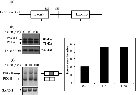
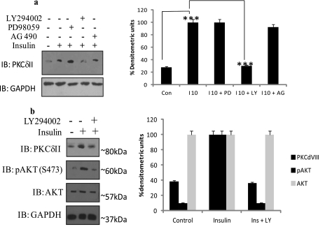
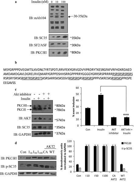
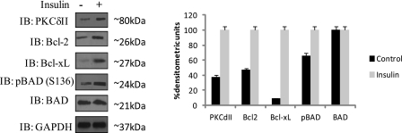
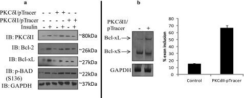
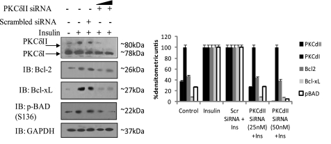
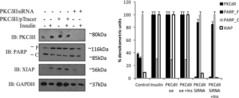
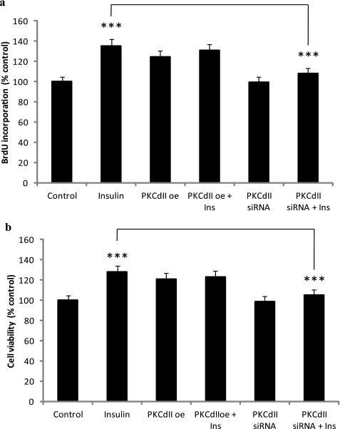
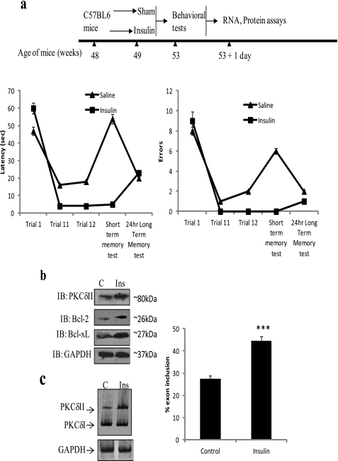
Insulin signaling pathways in the brain regulate food uptake and memory and learning. Insulin and protein kinase C (PKC) pathways are integrated and function closely together. PKC activation in the brain is essential for learning and neuronal repair. Intranasal delivery of insulin to the central nervous system (CNS) has been shown to improve memory, reduce cerebral atrophy, and reverse neurodegeneration. However, the neuronal molecular mechanisms of these effects have not been studied in depth. PKCδ plays a central role in cell survival. Its splice variants, PKCδI and PKCδII, are switches that determine cell survival and fate. PKCδI promotes apoptosis, whereas PKCδII promotes survival. Here, we demonstrate that insulin promotes alternative splicing of PKCδII isoform in HT22 cells. The expression of PKCδI splice variant remains unchanged. Insulin increases PKCδII alternative splicing via the PI3K pathway. We further demonstrate that Akt kinase mediates phosphorylation of the splicing factor SC35 to promote PKCδII alternative splicing. Using overexpression and knockdown assays, we demonstrate that insulin increases expression of Bcl2 and bcl-xL via PKCδII. We demonstrate increased cell proliferation and increased BrdU incorporation in insulin-treated cells as well as in HT22 cells overexpressing PKCδII. Finally, we demonstrate in vivo that intranasal insulin promotes cognitive function in mice with concomitant increases in PKCδII expression in the hippocampus. This is the first report of insulin, generally considered a growth or metabolic hormone, regulating the alternative isoform expression of a key signaling kinase in neuronal cells such that it results in increased neuronal survival.


Figures

FIGURE 1.
FIGURE 1.
Insulin increases PKCδII expression. HT22 cells were treated with increasing doses (10 and 100 nm) of insulin for 24 h. a, whole protein lysates were separated and analyzed by Western blot analysis using PKCδ antibody and anti-GAPDH. b, total RNA was extracted and RT-PCR performed with primers that simultaneously detect PKCδI and PKCδII mRNA. The gels are representative of four experiments performed with similar results. The graph represents the percentage of exon inclusion calculated as SS II/(SS II + SS I) × 100 and is representative of four experiments performed separately.
FIGURE 2.
FIGURE 2.
a, PI3K inhibitor LY294002 blocked insulin-mediated increase of PKCδII expression. HT22 cells were treated with signaling cascade inhibitors: PI3K inhibitor, LY294002 (1 μm), Janus-activated kinase inhibitor, AG490 (5 μm), or MEK inhibitor, PD98059 (10 μm) for 30 min prior to the addition of 10 nm insulin. Whole cell lysates were analyzed by Western blot analysis using antibodies as indicated. The graph shows PKCδII densitometric units normalized to GAPDH and represents three separate experiments. The results were analyzed with a two-tailed Student's t test using PRISM4 statistical analysis software (GraphPad, San Diego, CA). A level of p < 0.05 was considered statistically significant. ***, p < 0.0001. Significance was determined after three or more experiments. b, LY294002 blocked insulin-mediated increase of PKCδII expression and phosphorylation of Akt kinase. HT22 cells were treated with the PI3K inhibitor, LY294002 (1 μm), for 30 min prior to the addition of 10 nm insulin for 24 h. Whole cell lysates were analyzed by Western blot analysis using antibodies against PKCδII, p-AKT, AKT, and GAPDH as indicated. The gels represent three experiments performed separately with similar results. The graph shows PKCδII densitometric units normalized to GAPDH and represents three separate experiments. The results were analyzed with a two-tailed Student's t test using PRISM4 statistical analysis software (GraphPad). A level of p < 0.05 was considered statistically significant. ***, p < 0.0001. Significance was determined after three or more experiments.
FIGURE 3.
FIGURE 3.
a, insulin increases alternative splicing of PKCδII mRNA via SC35. HT22 cells were treated with increasing doses (10, 50, and 100 nm) of insulin for 24 h or left untreated (control). Whole protein lysates were separated and analyzed by Western blot analysis using either mAb104, PKCδII, SF2/ASF, or SC35antibody as indicated. The gels represent three experiments performed separately with similar results. b, SC35 protein sequence showing Akt kinase consensus sequences RXRXX(S/T) in its RS domain. c, HT22 cells were treated with the Akt inhibitor (124005, 1 μm) for 30 min prior to the addition of 10 nm insulin for 24 h. Whole cell lysates were analyzed by Western blot analysis using antibodies as indicated. The gels represent three experiments performed separately with similar results. The graph represents the percentage of exon inclusion calculated as SS II/(SS II + SS I) × 100 and is representative of four experiments performed separately. The results were analyzed with a two-tailed Student's t test using PRISM4 statistical analysis software (GraphPad). A level of p < 0.05 was considered statistically significant. ***, p < 0.0001. Significance was determined after three or more experiments. d, HT22 cells were treated with increasing doses (10, 50, and 100 nm) of insulin for 24 h or transiently transfected with CA-Akt2 or WT-Akt2. Whole protein lysates were separated and analyzed by Western blot analysis using either PKCδII, phospho-SC35, or GAPDH antibody as indicated. The gels represent three experiments performed separately with similar results.
FIGURE 4.
FIGURE 4.
Insulin increases expression of the pro-survival proteins. HT22 cells were treated with 10 nm insulin for 24 h. Whole cell lysates were analyzed by Western blot analysis using antibodies against PKCδII, Bcl2, bcl-xL, p-BAD, BAD, and GAPDH as indicated. The gels represent four experiments performed separately with similar results. The graph shows PKCδII densitometric units normalized to GAPDH and represents three separate experiments. The results were analyzed with a two-tailed Student's t test using PRISM4 statistical analysis software (GraphPad). A level of p < 0.05 was considered statistically significant. ***, p < 0.0001. Significance was determined after three or more experiments.
FIGURE 5.
FIGURE 5.
a, overexpression of PKCδII increased expression levels of Bcl2 and BclxL. HT22 cells were transfected with either PKCδI-pTracer or PKCδII-pTracer vector overnight. The cells were then treated with 10 nm insulin or left untreated for 24 h. Whole cell lysates were analyzed by Western blot analysis using antibodies against PKCδII, Bcl2, bcl-xL, p-BAD, and GAPDH as indicated. The gels represent three experiments performed separately with similar results. b, overexpression of PKCδII promotes Bcl-xL alternative splicing. HT22 cells were transfected with PKCδII-pTracer vector and RT-PCR performed with Bcl-x primers that detect Bcl-xS and Bcl-xL levels simultaneously. The graph represents the percentage of exon inclusion calculated as Bcl-xL/(Bcl-xS + Bcl-xL) × 100 and is representative of four experiments performed separately.
FIGURE 6.
FIGURE 6.
Knockdown of PKCδII decreased expression levels of Bcl2 and Bcl-xL. HT22 cells were transfected with increasing doses (25 and 50 nm) of PKCδII siRNA (indicated by the triangle above the lanes) or scrambled siRNA for 48 h. The cells were then treated with 10 nm insulin for 24 h. Whole cell lysates were analyzed by Western blot analysis using COOH-terminal PKCδ antibody which recognizes both PKCδI and -δII isoforms, anti-Bcl2, anti-Bcl-xL, anti-phosphoBAD, or anti-GAPDH as indicated. The gels represent three experiments performed separately with similar results. The graph shows the percentage of densitometric units normalized to GAPDH for each antibody and represents three separate experiments.
FIGURE 7.
FIGURE 7.
Insulin decreases neuronal apoptosis via PKCδII. HT22 cells were transfected with either 2 μg of PKCδII-pTracer vector or 50 nm PKCδII siRNA for 48 h and then were treated with 10 nm insulin overnight as indicated. Whole cell lysates were analyzed by Western blot analysis using antibodies against PKCδII, XIAP, or PARP. PARP_F, full-length PARP; PARP_C, cleaved fragment of PARP. The gels represent three experiments performed separately with similar results. The graph shows the percentage of densitometric units normalized to GAPDH for each antibody and represents three separate experiments.
FIGURE 8.
FIGURE 8.
Overexpression of PKCδII increases HT22 proliferation and viability. HT22 cells were transfected with either 2 μg of PKCδII-pTracer vector or 50 nm PKCδII siRNA for 48 h and then were treated with 10 nm insulin overnight as indicated. The BrdU assay and cell viability assay were performed. The graphs represent BrdU incorporation in PKCδII-overexpressing cells as a percentage of control cells (a) and cell viability in PKCδII-overexpressing cells as a percentage of control cells (b). The measurements were made in triplicate in three separate experiments. The results were analyzed with a two-tailed Student's t test using PRISM4 statistical analysis software (GraphPad). A level of p < 0.05 was considered statistically significant. ***, p < 0.0001. Experiments were performed in triplicate, and significance was determined after three experiments.
FIGURE 9.
FIGURE 9.
Intranasal insulin improved cognitive functions in vivo with concurrent increase in PKCδII expression. a, experimental setup and behavior tests after intranasal insulin treatment. Performance during the last block of pre-testing in the radial arm water maze task during working memory trials T11 and/or T12 is shown; retention trial (short term memory) and long term memory retention for both errors and latency measurements are shown. Results revealed insulin treatment improved performance in memory tests. The hippocampus was harvested and analyzed with Western blot analysis using PKCδII-specific antibody (b) and RT-PCR analysis using primers that simultaneously detect PKCδI and PKCδII mRNA levels (c). The gels are representative of six mice per treatment in experiments performed with similar results. The graph represents the percentage of exon inclusion calculated as SS II/(SS II + SS I) × 100 and is representative of six experiments performed separately. The results were analyzed with a two-tailed Student's t test using PRISM4 statistical analysis software (GraphPad). A level of p < 0.05 was considered statistically significant. ***, p < 0.0001.

Similar articles

Cited by

References

    1. Benedict C., Hallschmid M., Schmitz K., Schultes B., Ratter F., Fehm H. L., Born J., Kern W. (2007) Intranasal insulin improves memory in humans. Superiority of insulin aspart. Neuropsychopharmacology 32, 239–243 - PubMed
    1. Schmidt H., Kern W., Giese R., Hallschmid M., Enders A. (2009) Intranasal insulin to improve developmental delay in children with 22q13 deletion syndrome. An exploratory clinical trial. J. Med. Genet. 46, 217–222 - PubMed
    1. Park C. R., Seeley R. J., Craft S., Woods S. C. (2000) Intracerebroventricular insulin enhances memory in a passive-avoidance task. Physiol. Behav. 68, 509–514 - PubMed
    1. Francis G. J., Martinez J. A., Liu W. Q., Xu K., Ayer A., Fine J., Tuor U. I., Glazner G., Hanson L. R., Frey W. H., 2nd, Toth C. (2008) Intranasal insulin prevents cognitive decline, cerebral atrophy and white matter changes in murine type I diabetic encephalopathy. Brain 131, 3311–3334 - PubMed
    1. Francis G. J., Martinez J. A., Liu W. Q., Xu K., Ayer A., Fine J., Tuor U. I., Glazner G., Hanson L. R., Frey W. H., 2nd, Toth C. (2008) Intranasal insulin prevents cognitive decline, cerebral atrophy and white matter changes in murine type I diabetic encephalopathy. Brain 131, 3311–3334 - PubMed

Publication types

MeSH terms