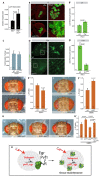
Schnurri regulates hemocyte function to promote tissue recovery after DNA damage
- PMID: 22275438
- PMCID: PMC3336376
- DOI: 10.1242/jcs.095323
Schnurri regulates hemocyte function to promote tissue recovery after DNA damage
Abstract
Tissue recovery after injury requires coordinated regulation of cell repair and apoptosis, removal of dead cells and regeneration. A critical step in this process is the recruitment of blood cells that mediate local inflammatory and immune responses, promoting tissue recovery. Here we identify a new role for the transcriptional regulator Schnurri (Shn) in the recovery of UV-damaged Drosophila retina. Using an experimental paradigm that allows precise quantification of tissue recovery after a defined dose of UV, we find that Shn activity in the retina is required to limit tissue damage. This function of Shn relies on its transcriptional induction of the PDGF-related growth factor Pvf1, which signals to tissue-associated hemocytes. We show that the Pvf1 receptor PVR acts in hemocytes to induce a macrophage-like morphology and that this is required to limit tissue loss after irradiation. Our results identify a new Shn-regulated paracrine signaling interaction between damaged retinal cells and hemocytes that ensures recovery and homeostasis of the challenged tissue.
Figures


Similar articles
-
Pvr and distinct downstream signaling factors are required for hemocyte spreading and epidermal wound closure at Drosophila larval wound sites.G3 (Bethesda). 2022 Jan 4;12(1):jkab388. doi: 10.1093/g3journal/jkab388. G3 (Bethesda). 2022. PMID: 34751396 Free PMC article.
-
Murine Schnurri-2 is required for positive selection of thymocytes.Nat Immunol. 2001 Nov;2(11):1048-53. doi: 10.1038/ni728. Nat Immunol. 2001. PMID: 11668343
-
Dmp53 protects the Drosophila retina during a developmentally regulated DNA damage response.EMBO J. 2003 Oct 15;22(20):5622-32. doi: 10.1093/emboj/cdg543. EMBO J. 2003. PMID: 14532134 Free PMC article.
-
Thicker than blood: conserved mechanisms in Drosophila and vertebrate hematopoiesis.Dev Cell. 2003 Nov;5(5):673-90. doi: 10.1016/s1534-5807(03)00335-6. Dev Cell. 2003. PMID: 14602069 Review.
-
The roles of fruitless and doublesex in the control of male courtship.Int Rev Neurobiol. 2011;99:87-105. doi: 10.1016/B978-0-12-387003-2.00004-5. Int Rev Neurobiol. 2011. PMID: 21906537 Review.
Cited by
-
Single-cell RNA-seq revealed heterogeneous responses and functional differentiation of hemocytes against white spot syndrome virus infection in Litopenaeus vannamei.J Virol. 2024 Mar 19;98(3):e0180523. doi: 10.1128/jvi.01805-23. Epub 2024 Feb 7. J Virol. 2024. PMID: 38323810 Free PMC article.
-
Vertebrate-conserved Schnurri zinc fingers restrain Drosophila vein patterning.MicroPubl Biol. 2024 Jan 26;2024:10.17912/micropub.biology.001066. doi: 10.17912/micropub.biology.001066. eCollection 2024. MicroPubl Biol. 2024. PMID: 38344065 Free PMC article.
-
What Drosophila Can Teach Us About Radiation Biology of Human Cancers.Adv Exp Med Biol. 2019;1167:225-236. doi: 10.1007/978-3-030-23629-8_13. Adv Exp Med Biol. 2019. PMID: 31520358 Review.
-
Dpp/TGFβ-superfamily play a dual conserved role in mediating the damage response in the retina.PLoS One. 2021 Oct 26;16(10):e0258872. doi: 10.1371/journal.pone.0258872. eCollection 2021. PLoS One. 2021. PMID: 34699550 Free PMC article.
-
Macrophages and cellular immunity in Drosophila melanogaster.Semin Immunol. 2015 Dec;27(6):357-68. doi: 10.1016/j.smim.2016.03.010. Epub 2016 Apr 23. Semin Immunol. 2015. PMID: 27117654 Free PMC article. Review.
References
-
- Arora K., Dai H., Kazuko S. G., Jamal J., O'Connor M. B., Letsou A., Warrior R. (1995). The Drosophila schnurri gene acts in the Dpp/TGF-beta signaling pathway and encodes a transcription factor homologous to the human MBP family. Cell 81, 781-790 - PubMed
-
- Bidla G., Dushay M. S., Theopold U. (2007). Crystal cell rupture after injury in Drosophila requires the JNK pathway, small GTPases and the TNF homolog Eiger. J. Cell Sci. 120, 1209-1215 - PubMed
-
- Brückner K., Kockel L., Duchek P., Luque C. M., Rørth P., Perrimon N. (2004). The PDGF/VEGF receptor controls blood cell survival in Drosophila. Dev. Cell 7, 73-84 - PubMed
Publication types
MeSH terms
Substances
Grants and funding
LinkOut - more resources
Full Text Sources
Molecular Biology Databases
Research Materials

