. 2012 Feb 17;110(4):598-608.
doi: 10.1161/CIRCRESAHA.111.258285. Epub 2012 Jan 24.

Hyperamylinemia contributes to cardiac dysfunction in obesity and diabetes: a study in humans and rats

Affiliations

Hyperamylinemia contributes to cardiac dysfunction in obesity and diabetes: a study in humans and rats

Sanda Despa et al. Circ Res. .

Abstract

Rationale: Hyperamylinemia is common in patients with obesity and insulin resistance, coincides with hyperinsulinemia, and results in amyloid deposition. Amylin amyloids are generally considered a pancreatic disorder in type 2 diabetes. However, elevated circulating levels of amylin may also lead to amylin accumulation and proteotoxicity in peripheral organs, including the heart.

Objective: To test whether amylin accumulates in the heart of obese and type 2 diabetic patients and to uncover the effects of amylin accumulation on cardiac morphology and function.

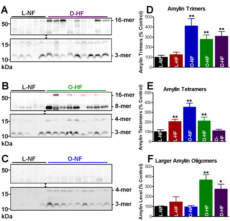
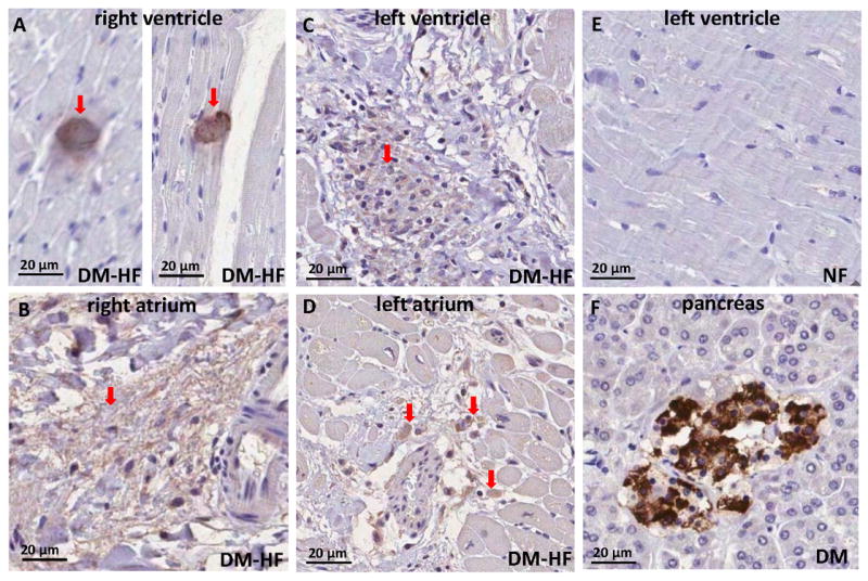
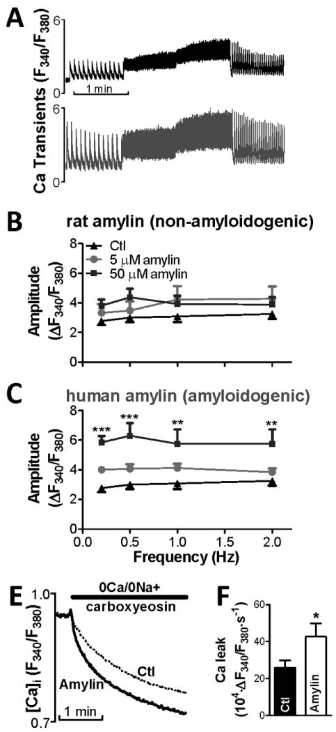
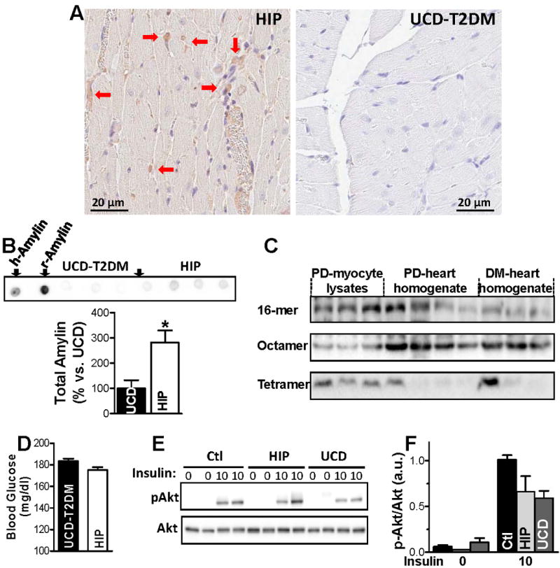
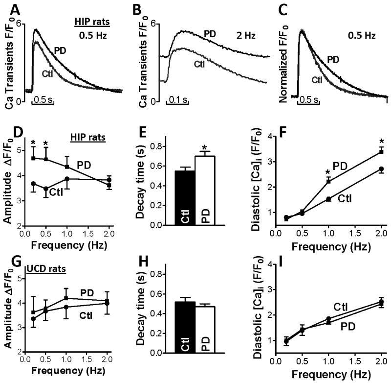
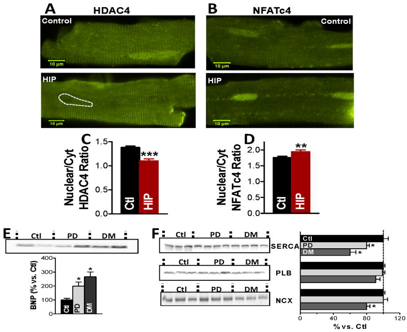
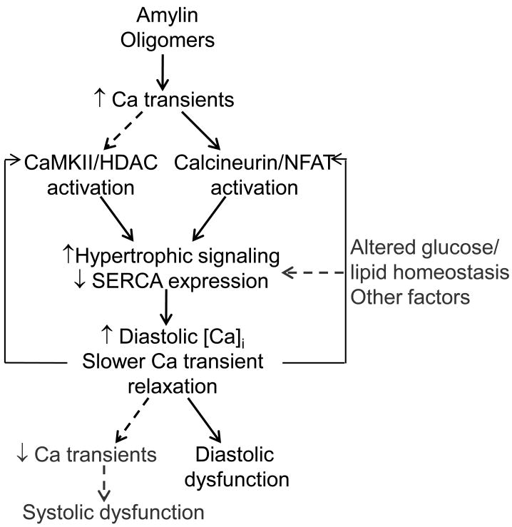
Methods and results: We compared amylin deposition in failing and nonfailing hearts from lean, obese, and type 2 diabetic humans using immunohistochemistry and Western blots. We found significant accumulation of large amylin oligomers, fibrils, and plaques in failing hearts from obese and diabetic patients but not in normal hearts and failing hearts from lean, nondiabetic humans. Small amylin oligomers were even elevated in nonfailing hearts from overweight/obese patients, suggesting an early state of accumulation. Using a rat model of hyperamylinemia transgenic for human amylin, we observed that amylin oligomers attach to the sarcolemma, leading to myocyte Ca(2+) dysregulation, pathological myocyte remodeling, and diastolic dysfunction, starting from prediabetes. In contrast, prediabetic rats expressing the same level of wild-type rat amylin, a nonamyloidogenic isoform, exhibited normal heart structure and function.

Conclusions: Hyperamylinemia promotes amylin deposition in the heart, causing alterations of cardiac myocyte structure and function. We propose that detection and disruption of cardiac amylin buildup may be both a predictor of heart dysfunction and a novel therapeutic strategy in diabetic cardiomyopathy.


Figures

Figure 1
Figure 1
(A-C) Amylin oligomer size distribution in failing (HF) and non-failing (NF) hearts from diabetic (D) and overweight/obese (O) patients vs. lean (L) controls. Representative (of ≥4 experiments) western blots with anti-amylin antibody of left ventricle protein homogenates from hearts in the D-HF (A), O-HF (B) and O-NF (C) groups. In each case, comparisons are made with hearts from the L-NF group. Specific molecular weight bands correspond to amylin trimers (12 kDa), tetramers (16 kDa) and two larger molecular weight structures at ∼32 kDa (octamers) and ∼64 kDa (16-mers). (D-F) Intensity signal analysis of the 12, 16 and the integrated 32-64 kDa bands. Heart samples in the D-HF (N=25), O-HF (N=8) and O-NF (N=8) groups contain markedly higher amylin oligomer levels than the controls L-NF (N=5) and L-HF (N=7). Large size amylin octamers and 16-mers are abundantly present only in the failing heart O-HF and D-HF groups. *P < 0.05; **P < 0.01.
Figure 2
Figure 2
Amylin deposition in failing diabetic hearts demonstrated by immunohistochemistry with an anti-amylin antibody on thin heart sections. Amylin plaques (A,C, and D, arrows) and tangles (B, arrow) are scattered through the entire heart. (E) Left ventricle section from a non-failing heart; no amylin deposits are revealed. (F) Positive control for amylin deposition in a pancreas from a diabetic patient.
Figure 3
Figure 3
Exogenous human amylin oligomers increase Ca2+ transient amplitude in isolated rat cardiac myocytes. (A) Representative Ca2+ transient measurements in a control (top panel) cell and a myocyte pre-incubated with 50 μmol/L human amylin (bottom panel). (B-C) Effect of 5 and 50 μmol/L of rat (B) and human (C) amylin on Ca2+ transient amplitude. Oligomerization of human amylin resulted in a marked Ca2+ transient increase. (D) Passive sarcolemmal Ca2+ leak measurements as initial slope of [Ca2+]i decline upon reducing external [Ca2+] from 1 mmol/L to 0, with the SR, Na/Ca exchanger and sarcolemmal Ca2+-ATPase blocked by pre-treatment with thapsigargin, 0Na+/0Ca+ solution and 20 μmol/L carboxyeosin, respectively. (E) Passive trans-sarcolemmal Ca2+ leak is significantly larger in myocytes pre-treated with 50 μmol/L human amylin vs. control. For each group, measurements were done on ≥6 myocytes from 3 different rats.
Figure 4
Figure 4
(A) Immunohistochemistry with an anti-amylin antibody on thin heart sections demonstrating amylin deposition in cardiac tissues from pre-diabetic HIP but not UCD-T2DM rats (20X). (B) Dot blots with the anti-amylin antibody comparing total amylin level in HIP vs. UCD-T2DM rats. The first two dots on the left show positive controls using 5 ng of recombinant human (h-Amylin) and rat (r-Amylin) amylin. The antibody binds r-Amylin with significantly higher affinity than h-amylin; The bottom panel shows the average signal intensity in hearts from pre-diabetic HIP vs. UCD-T2DM rats. The experiment was performed in triplicate. (C) Representative western blot with anti-amylin primary antibody on ventricular myocyte lysates from pre-diabetic HIP rats, and left ventricle protein homogenates from pre-diabetic (PD) and diabetic (DM) HIP rats. (D) Blood glucose levels in age-matched pre-diabetic HIP rats (N=14) and UCD-T2DM rats (N=16). (E-F) Akt phosphorylation in hearts from pre-diabetic HIP and UCD-T2DM rats and littermate controls under basal conditions (0 insulin) and following stimulation with insulin (10 mU/g body weight). Representative example (E) and mean values for the ratio between phosphorylated and total Akt (F). N=3 rats for each group.
Figure 5
Figure 5
Altered Ca2+ cycling in cardiac myocytes from pre-diabetic HIP but not pre-diabetic UCD-T2DM rats. Representative Ca2+ transients in myocytes from control (Ctl) and pre-diabetic (PD) HIP rats paced at 0.5 Hz (A) and 2 Hz (B). (C) Normalized Ca2+ transients in myocytes from control and pre-diabetic HIP rats (0.5 Hz) indicate slower Ca2+ transient relaxation in pre-diabetic HIP rats vs. control. (D) Mean amplitude of Ca2+ transients recorded in cardiac myocytes from control rats (20 myocytes, 4 rats) and pre-diabetic HIP rats (18 cells, 4 rats) paced at 0.2, 0.5, 1 and 2 Hz. At 0.2 and 0.5 Hz, Ca2+ transient amplitude is significantly larger in myocytes from pre-diabetic HIP rats vs. control. This difference disappears at higher stimulation frequencies. (E) Ca2+ transient decay time in cardiac myocytes from control and pre-diabetic HIP rats paced at 0.5 Hz. (F) Diastolic [Ca2+]i in cardiac myocytes from control rats and pre-diabetic HIP rats paced at 0.2, 0.5, 1 and 2 Hz. At higher frequencies, diastolic [Ca2+]i is significantly higher in myocytes from pre-diabetic HIP vs. control rats. (G) Mean amplitude of Ca2+ transients in myocytes from control rats (22 myocytes, 6 rats) and pre-diabetic UCD-T2DM rats (21 cells, 4 rats) paced at 0.2, 0.5, 1 and 2 Hz. (H) Ca transient decay time in myocytes from control and pre-diabetic UCD-T2DM rats paced at 0.5 Hz. (I) Diastolic [Ca2+]i in myocytes from control and pre-diabetic UCD-T2DM rats paced at 0.2, 0.5, 1 and 2 Hz. *P<0.05
Figure 6
Figure 6
HDAC4 nuclear export, NFATc4 nuclear import, reduced SERCA and increased BNP in hearts from pre-diabetic HIP rats. (A-B) Representative immunofluorescence images showing the distribution of HDAC4 and, respectively, NFATc4 in myocytes from pre-diabetic HIP rats and age-matched WT rats. (C-D) The nuclear-to-cytosolic ratio of HDAC4 (C) and NFATc4 (D) demonstrates the nuclear export of HDAC4 and nuclear import of NFATc4 in pre-diabetic HIP rats. Experiments were done on more than 15 cells from 3 rats for both groups. (E) Increased expression of the hyperthrophic marker BNP in hearts from pre-diabetic (PD) and diabetic (DM) HIP rats. (F) Alterations in the protein expression of SERCA, phospholamban and Na/Ca exchanger in hearts from pre-diabetic and diabetic HIP rats vs. control, non-diabetic rats (Ctl). Ctl – 5 hearts; PD – 5 hearts, DM – 5 hearts.
Figure 7
Figure 7
Proposed mechanism for amylin oligomer-induced cardiac dysfunction. Amylin oligomers elevate Ca2+ transients, which results in activation of Ca2+-dependent CaMKII-HDAC and calcineurin-NFAT transcriptional regulation/hypertrophic pathways. This may reduce SERCA expression, which further alters myocyte Ca2+ cycling by impairing Ca2+ transient relaxation leading to higher diastolic [Ca2+]i. Impaired Ca2+ transient relaxation and elevated diastolic [Ca2+]i may further activate the Ca2+-dependent transcriptional regulation/hypertrophic pathways (positive feedback) and cause diastolic dysfunction in HIP rats. With the advancement of the disease, reduced SERCA function may cause SR unloading, consequent reduction in Ca2+ transient amplitude, and systolic dysfunction.

References

    1. Biddinger SB, Kahn CR. From mice to men: insights into the insulin resistance syndromes. Annu Rev Physiol. 2006;68:123–158. - PubMed
    1. Lopaschuk GD, Ussher JR, Folmes CD, Jaswal JS, Stanley WC. Myocardial fatty acid metabolism in health and disease. Physiol Rev. 2010;90:207–258. - PubMed
    1. Reaven GM. Relationships among insulin resistance, type 2 diabetes, essential hypertension, and cardiovascular disease: similarities and differences. J Clin Hypertens. 2011;13:238–243. - PMC - PubMed
    1. Battiprolu PK, Gillette TG, Wang ZV, Lavandero S, Hill JA. Diabetic Cardiomyopathy: Mechanisms and Therapeutic Targets. Drug Discov Today Dis Mech. 2010;7:e135–e143. - PMC - PubMed
    1. Guha A, Harmancey R, Taegtmeyer H. Nonischemic heart failure in diabetes mellitus. Curr Opin Cardiol. 2008;23:241–248. - PMC - PubMed

Publication types

MeSH terms

Substances