. 2012 Mar 2;110(5):701-15.
doi: 10.1161/CIRCRESAHA.111.259507. Epub 2012 Jan 24.

Cardiomyogenesis in the developing heart is regulated by c-kit-positive cardiac stem cells

Affiliations

Cardiomyogenesis in the developing heart is regulated by c-kit-positive cardiac stem cells

João Ferreira-Martins et al. Circ Res. .

Retraction in

Expression of concern in

  • Expression of Concern.
    [No authors listed] [No authors listed] Circ Res. 2019 Jan 18;124(2):e4-e5. doi: 10.1161/RES.0000000000000241. Circ Res. 2019. PMID: 30582460 No abstract available.
  • Expression of Concern.
    [No authors listed] [No authors listed] Circulation. 2019 Jan 15;139(3):e5-e6. doi: 10.1161/CIR.0000000000000639. Circulation. 2019. PMID: 30615475 No abstract available.

Abstract

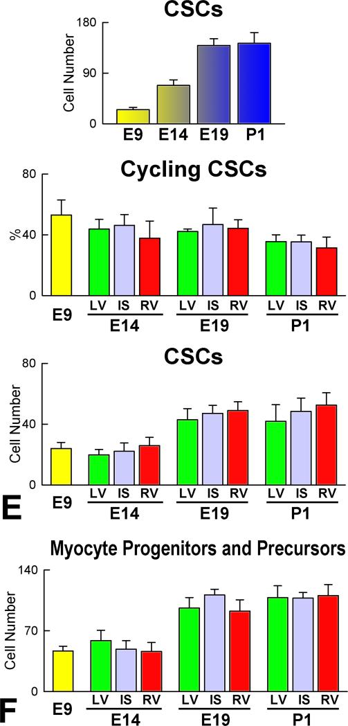
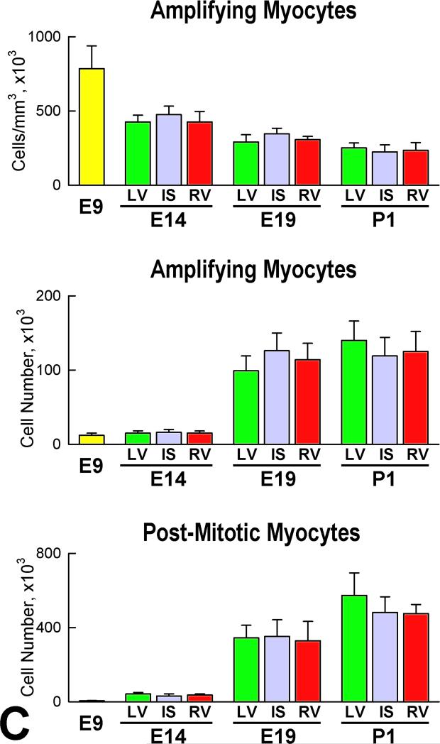
Rationale: Embryonic and fetal myocardial growth is characterized by a dramatic increase in myocyte number, but whether the expansion of the myocyte compartment is dictated by activation and commitment of resident cardiac stem cells (CSCs), division of immature myocytes or both is currently unknown.

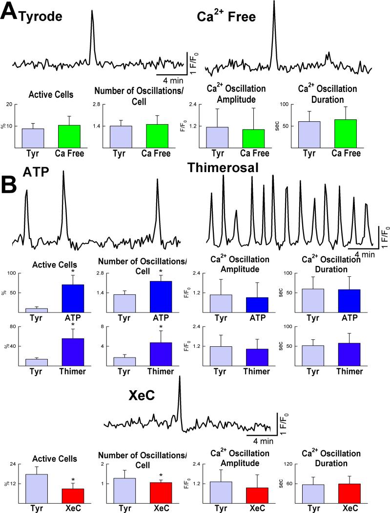
Objective: In this study, we tested whether prenatal cardiac development is controlled by activation and differentiation of CSCs and whether division of c-kit-positive CSCs in the mouse heart is triggered by spontaneous Ca(2+) oscillations.

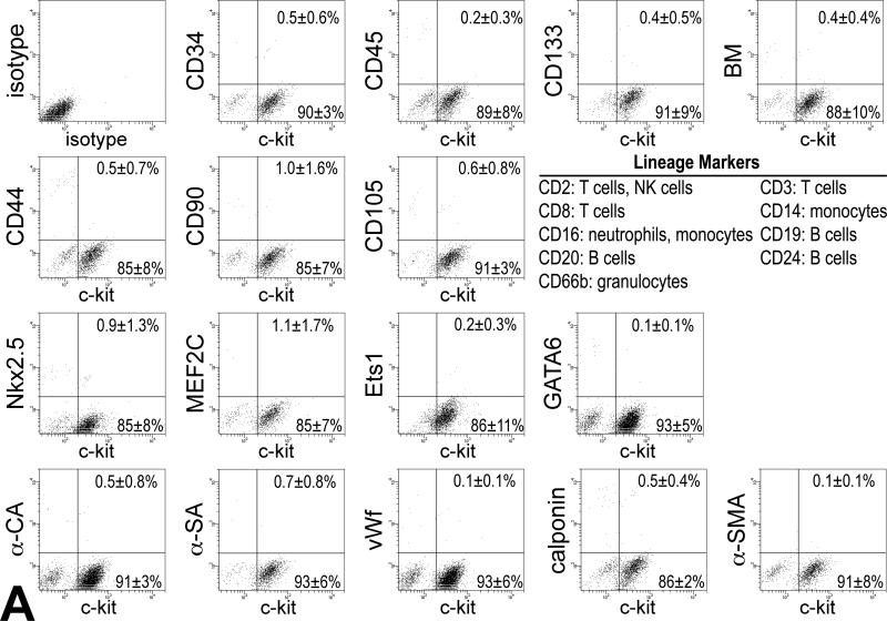
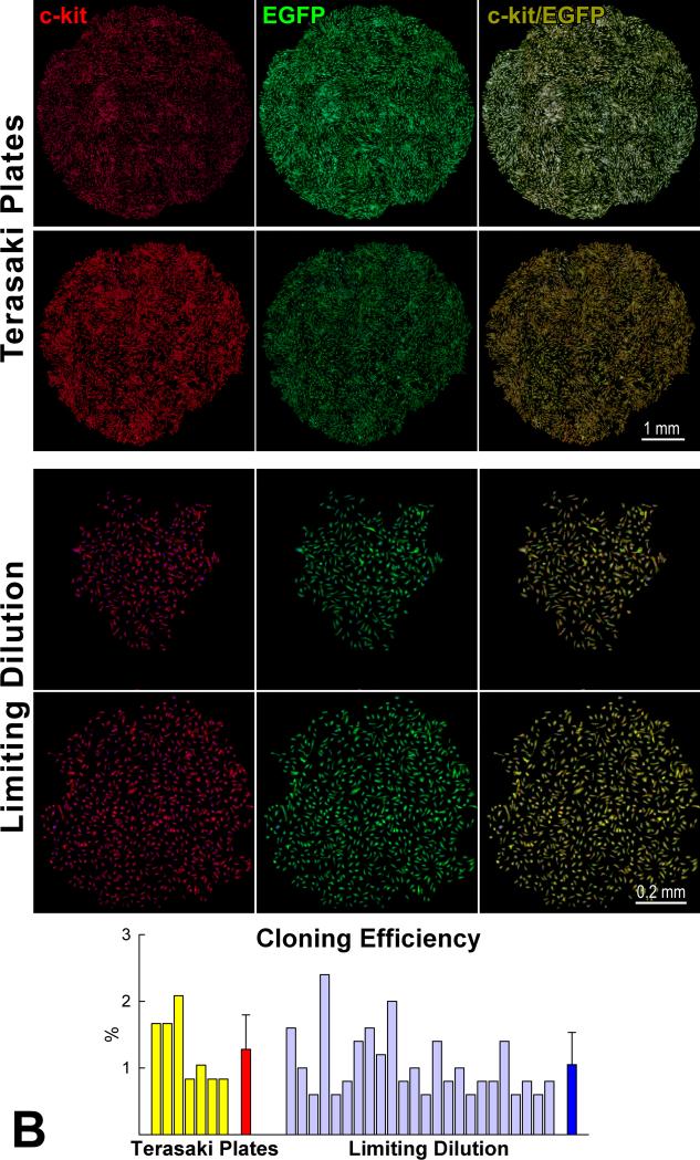
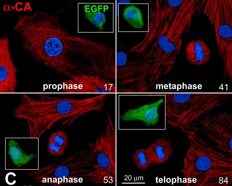
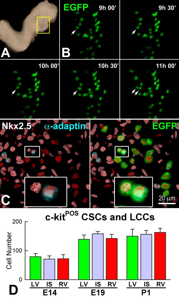
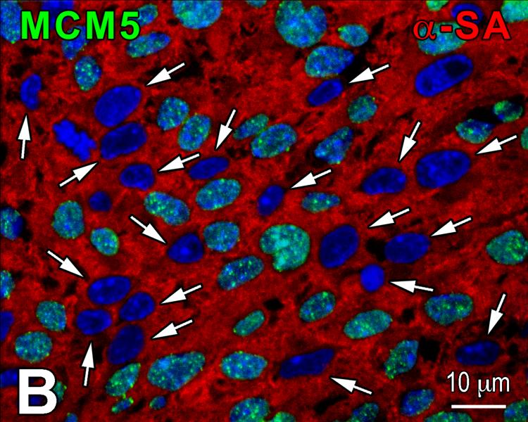
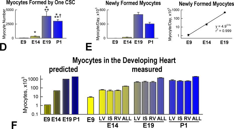
Methods and results: We report that embryonic-fetal c-kit-positive CSCs are self-renewing, clonogenic and multipotent in vitro and in vivo. The growth and commitment of c-kit-positive CSCs is responsible for the generation of the myocyte progeny of the developing heart. The close correspondence between values computed by mathematical modeling and direct measurements of myocyte number at E9, E14, E19 and 1 day after birth strongly suggests that the organogenesis of the embryonic heart is dependent on a hierarchical model of cell differentiation regulated by resident CSCs. The growth promoting effects of c-kit-positive CSCs are triggered by spontaneous oscillations in intracellular Ca(2+), mediated by IP3 receptor activation, which condition asymmetrical stem cell division and myocyte lineage specification.

Conclusions: Myocyte formation derived from CSC differentiation is the major determinant of cardiac growth during development. Division of c-kit-positive CSCs in the mouse is promoted by spontaneous Ca(2+) spikes, which dictate the pattern of stem cell replication and the generation of a myocyte progeny at all phases of prenatal life and up to one day after birth.


Figures

Figure 1
Figure 1
Co-expression of c-kit and EGFP. A: Bivariate distribution of c-kit and EGFP expression in small cardiac cells isolated at E18. B: By confocal microscopy, EGFP-positive cells at E15 (left panel, green) are positive for c-kit (central panel, red). Right panel, merge. C: Amplifying myocytes at E15 are EGFP-positive (left panel, green), and express Ki67 (central panel, magenta) and α-sarcomeric actin (central panel: α-SA, white). Right panel, merge. D: Longitudinal section of a c-kit-EGFP embryo at E6.5 shown by two-photon microscopy. Nuclei are stained by DAPI (left panel, blue). Clusters of pCSCs are present (central panel: EGFP, green, arrows). Right panel, merge. E: As shown by confocal microscopy, the cardiogenic mesoderm of a c-kit-EGFP embryo at E6.5 contains a group of c-kit-positive pCSCs (red). F through K: pCSC niches at E11.5. The heart of a c-kit-EGFP embryo is included in the area defined by a rectangle in F and G. H through K are higher magnifications by two-photon microscopy of the areas delineated by small squares in G; EGFP-positive pCSCs (green) are scattered in the myocardium of the common ventricular chamber (CVC, H and I) and are clustered at the apex (apex, J). K is a higher magnification by two-photon microscopy of the apical cluster of pCSCs in J. EGFP-positive pCSCs (green) express N-cadherin (white) at the interface with myocytes (α-cardiac actinin, α-CA, red). Nuclei: DAPI, blue.
Figure 1
Figure 1
Co-expression of c-kit and EGFP. A: Bivariate distribution of c-kit and EGFP expression in small cardiac cells isolated at E18. B: By confocal microscopy, EGFP-positive cells at E15 (left panel, green) are positive for c-kit (central panel, red). Right panel, merge. C: Amplifying myocytes at E15 are EGFP-positive (left panel, green), and express Ki67 (central panel, magenta) and α-sarcomeric actin (central panel: α-SA, white). Right panel, merge. D: Longitudinal section of a c-kit-EGFP embryo at E6.5 shown by two-photon microscopy. Nuclei are stained by DAPI (left panel, blue). Clusters of pCSCs are present (central panel: EGFP, green, arrows). Right panel, merge. E: As shown by confocal microscopy, the cardiogenic mesoderm of a c-kit-EGFP embryo at E6.5 contains a group of c-kit-positive pCSCs (red). F through K: pCSC niches at E11.5. The heart of a c-kit-EGFP embryo is included in the area defined by a rectangle in F and G. H through K are higher magnifications by two-photon microscopy of the areas delineated by small squares in G; EGFP-positive pCSCs (green) are scattered in the myocardium of the common ventricular chamber (CVC, H and I) and are clustered at the apex (apex, J). K is a higher magnification by two-photon microscopy of the apical cluster of pCSCs in J. EGFP-positive pCSCs (green) express N-cadherin (white) at the interface with myocytes (α-cardiac actinin, α-CA, red). Nuclei: DAPI, blue.
Figure 2
Figure 2
Morphogenetic movements of pCSCs. A through D: c-kit-EGFP embryo at E8.0 examined by two-photon microscopy for 12 hours. Images were recorded every 30 minutes; the 3 panels correspond to time 0 (A), 6 (B) and 12 (C) hours, respectively. The yellow oval delimits the heart and the red oval defines the yolk sac. During 12 hours, none of the EGFP-positive cells of the yolk sac (inside the red oval) migrated to the cardiac area (inside the yellow oval). However, pCSCs moved within the cardiac area (inside the yellow oval). Nuclei are stained by DAPI (blue) and cardiomyocytes by α-cardiac actinin (D: α-CA, red). E through J: c-kit-EGFP embryo at E8.5: the looping heart is surrounded by the oval (E). The square defines the portion of the embryo examined by two-photon microscopy for 5 hours. Images were recorded hourly: baseline (F), 2 (G) and 5 (H) hours. The yellow oval delimits the heart. During 5 hours, none of the EGFP-positive cells outside the heart (outside the oval) migrated to the cardiac area (inside the oval). I and J: The location of the heart was confirmed by myocyte labeling (α-CA, red) at the end of the experiment. EGFP-positive cells are located within the myocardium.
Figure 2
Figure 2
Morphogenetic movements of pCSCs. A through D: c-kit-EGFP embryo at E8.0 examined by two-photon microscopy for 12 hours. Images were recorded every 30 minutes; the 3 panels correspond to time 0 (A), 6 (B) and 12 (C) hours, respectively. The yellow oval delimits the heart and the red oval defines the yolk sac. During 12 hours, none of the EGFP-positive cells of the yolk sac (inside the red oval) migrated to the cardiac area (inside the yellow oval). However, pCSCs moved within the cardiac area (inside the yellow oval). Nuclei are stained by DAPI (blue) and cardiomyocytes by α-cardiac actinin (D: α-CA, red). E through J: c-kit-EGFP embryo at E8.5: the looping heart is surrounded by the oval (E). The square defines the portion of the embryo examined by two-photon microscopy for 5 hours. Images were recorded hourly: baseline (F), 2 (G) and 5 (H) hours. The yellow oval delimits the heart. During 5 hours, none of the EGFP-positive cells outside the heart (outside the oval) migrated to the cardiac area (inside the oval). I and J: The location of the heart was confirmed by myocyte labeling (α-CA, red) at the end of the experiment. EGFP-positive cells are located within the myocardium.
Figure 3
Figure 3
Characteristics of pCSCs. A: pCSCs were predominantly negative for markers of bone marrow cells, mesenchymal stromal cells, cardiomyocytes, ECs and SMCs. B: Four single cell-derived clones are shown (Terasaki plates: upper two; limiting dilution: lower two). Cells in the clones expressed c-kit (left, red) and EGFP (central, green). Right panel, merge. Cloning efficiency in each experiment is shown together with mean±SD. C: Dividing fetal myocytes in culture do not express EGFP. The number in each panel reflects sequential sampling of 102 cells. EGFP-positive CSCs (insets, green), positive control. α-CA, red. Chromosomes are stained by propidium iodide (blue).
Figure 3
Figure 3
Characteristics of pCSCs. A: pCSCs were predominantly negative for markers of bone marrow cells, mesenchymal stromal cells, cardiomyocytes, ECs and SMCs. B: Four single cell-derived clones are shown (Terasaki plates: upper two; limiting dilution: lower two). Cells in the clones expressed c-kit (left, red) and EGFP (central, green). Right panel, merge. Cloning efficiency in each experiment is shown together with mean±SD. C: Dividing fetal myocytes in culture do not express EGFP. The number in each panel reflects sequential sampling of 102 cells. EGFP-positive CSCs (insets, green), positive control. α-CA, red. Chromosomes are stained by propidium iodide (blue).
Figure 4
Figure 4
Ca2+ oscillations in pCSCs. A: Tracings of cytosolic Ca2+ in embryonic pCSCs exposed to Tyrode solution (Tyr) and Ca2+-free medium. B: Frequency, amplitude and duration of Ca2+ oscillations after activation of IP3 receptors by ATP and thimerosal (Thimer), or their inhibition by xestospongin-C (XeC). *P<0.05 vs. Tyr. C: Ca2+ oscillations in pCSCs treated with short hairpin-RNA (sh-RNA) targeting IP3 receptor type-2. *,†P<0.05 vs. control conditions (Ctrl) and Tyr, respectively. D: Changes in ryanodine receptor function by caffeine and ryanodine and Ca2+ oscillations in pCSCs.
Figure 4
Figure 4
Ca2+ oscillations in pCSCs. A: Tracings of cytosolic Ca2+ in embryonic pCSCs exposed to Tyrode solution (Tyr) and Ca2+-free medium. B: Frequency, amplitude and duration of Ca2+ oscillations after activation of IP3 receptors by ATP and thimerosal (Thimer), or their inhibition by xestospongin-C (XeC). *P<0.05 vs. Tyr. C: Ca2+ oscillations in pCSCs treated with short hairpin-RNA (sh-RNA) targeting IP3 receptor type-2. *,†P<0.05 vs. control conditions (Ctrl) and Tyr, respectively. D: Changes in ryanodine receptor function by caffeine and ryanodine and Ca2+ oscillations in pCSCs.
Figure 5
Figure 5
Ca2+ oscillations and division of pCSCs A: Duration of the phases of the cell cycle in embryonic pCSCs. B: DNA content in embryonic pCSCs before synchronization, and 1 and 8 hours after cell synchronization in mitosis. C: Ca2+ oscillations in synchronized pCSCs during the different phases of the cell cycle. *,†P<0.05 vs. G1 and S, respectively. D: pCSCs synchronized in G1: Ca2+ oscillations induced by ATP favor cell replication, while inhibition of Ca2+ oscillations by U73122 and xestospongin-C (XeC), or reduced expression of IP3 receptor type-2 by sh-RNA have opposite effects. *,†P<0.05 vs. Ctrl and ATP alone, respectively. Baseline condition (Base): pCSCs infected with control vector. E: Symmetric (left) and asymmetric (central and right) division of pCSCs. Asymmetric division (right) results in the expression of Nkx2.5 in one of the two daughter cells. F: IP3 receptor function modulates the fate of dividing pCSCs. *,†P<0.05 vs. symmetric division (S) and Ctrl, respectively. A, asymmetric division.
Figure 5
Figure 5
Ca2+ oscillations and division of pCSCs A: Duration of the phases of the cell cycle in embryonic pCSCs. B: DNA content in embryonic pCSCs before synchronization, and 1 and 8 hours after cell synchronization in mitosis. C: Ca2+ oscillations in synchronized pCSCs during the different phases of the cell cycle. *,†P<0.05 vs. G1 and S, respectively. D: pCSCs synchronized in G1: Ca2+ oscillations induced by ATP favor cell replication, while inhibition of Ca2+ oscillations by U73122 and xestospongin-C (XeC), or reduced expression of IP3 receptor type-2 by sh-RNA have opposite effects. *,†P<0.05 vs. Ctrl and ATP alone, respectively. Baseline condition (Base): pCSCs infected with control vector. E: Symmetric (left) and asymmetric (central and right) division of pCSCs. Asymmetric division (right) results in the expression of Nkx2.5 in one of the two daughter cells. F: IP3 receptor function modulates the fate of dividing pCSCs. *,†P<0.05 vs. symmetric division (S) and Ctrl, respectively. A, asymmetric division.
Figure 6
Figure 6
Transplantation assay. A: Clone of c-kit-positive CSCs (left, green), labeled by RFP (central, red). Right, merge. B: At 48 hours, transplanted RFP-positive pCSCs (red) are engrafted and express connexin-43 (left: Cx43, yellow, arrows) at the interface with recipient myocytes (α-SA, white). The expression of Nkx2.5 is also apparent (right: green, arrows). C: Dividing clonal CSCs with nuclei in telophase (left, green); the unipolar localization of α-adaptin (central, bright blue) documents asymmetric division. The expression of Nkx2.5 (white), Ets1 (yellow), and GATA6 (magenta) in one of the two daughter cells (right) illustrates the acquisition of the myocyte, EC, and SMC lineage, respectively. D: At 10 days, regenerated myocytes are positive for α-SA (upper, white), EGFP (central, green), and RFP (lower, red). Insets show at higher magnification the area included in the rectangle. E: EGFP-positive (left, green), α-SA-positive (right, white) regenerated myocytes display sarcomere striation (arrowheads).
Figure 6
Figure 6
Transplantation assay. A: Clone of c-kit-positive CSCs (left, green), labeled by RFP (central, red). Right, merge. B: At 48 hours, transplanted RFP-positive pCSCs (red) are engrafted and express connexin-43 (left: Cx43, yellow, arrows) at the interface with recipient myocytes (α-SA, white). The expression of Nkx2.5 is also apparent (right: green, arrows). C: Dividing clonal CSCs with nuclei in telophase (left, green); the unipolar localization of α-adaptin (central, bright blue) documents asymmetric division. The expression of Nkx2.5 (white), Ets1 (yellow), and GATA6 (magenta) in one of the two daughter cells (right) illustrates the acquisition of the myocyte, EC, and SMC lineage, respectively. D: At 10 days, regenerated myocytes are positive for α-SA (upper, white), EGFP (central, green), and RFP (lower, red). Insets show at higher magnification the area included in the rectangle. E: EGFP-positive (left, green), α-SA-positive (right, white) regenerated myocytes display sarcomere striation (arrowheads).
Figure 7
Figure 7
Asymmetric division and lineage specification of CSCs. A: The heart tube in a c-kit-EGFP mouse Embryo at E8 is defined by a yellow rectangle. This region was examined by two-photon microscopy for 11 hours. B: Within the heart tube, one EGFP-positive CSC underwent complete division from 9:30 to 11 hours. C: The divided cell was then analyzed by whole-mount immunolabeling and confocal microscopy. Left: one of the two daughter cells was positive for α-adaptin (blue) while the other expressed Nkx2.5 (white). Labeling for α-adaptin and Nkx2.5 in the dividing CSC is shown at higher magnification in the insets. Right: Labeling of EGFP in the dividing CSC. EGFP localization is shown at higher magnification in the inset. D: Aggregate number of c-kit-positive CSCs and lineage committed cells (LCCs: myocyte progenitors-precursors) in LV, IS, and RV at E14, E19 and P1. E and F: Number of c-kit-positive lineage-negative CSCs (E), and myocyte progenitors-precursors (F) in the entire heart, and in LV, IS and RV at E9, E14, E19 and P1.
Figure 7
Figure 7
Asymmetric division and lineage specification of CSCs. A: The heart tube in a c-kit-EGFP mouse Embryo at E8 is defined by a yellow rectangle. This region was examined by two-photon microscopy for 11 hours. B: Within the heart tube, one EGFP-positive CSC underwent complete division from 9:30 to 11 hours. C: The divided cell was then analyzed by whole-mount immunolabeling and confocal microscopy. Left: one of the two daughter cells was positive for α-adaptin (blue) while the other expressed Nkx2.5 (white). Labeling for α-adaptin and Nkx2.5 in the dividing CSC is shown at higher magnification in the insets. Right: Labeling of EGFP in the dividing CSC. EGFP localization is shown at higher magnification in the inset. D: Aggregate number of c-kit-positive CSCs and lineage committed cells (LCCs: myocyte progenitors-precursors) in LV, IS, and RV at E14, E19 and P1. E and F: Number of c-kit-positive lineage-negative CSCs (E), and myocyte progenitors-precursors (F) in the entire heart, and in LV, IS and RV at E9, E14, E19 and P1.
Figure 8
Figure 8
Myocyte formation. A: Amplifying myocytes (α-SA, red) are positive for MCM5 (left: green, arrows), phospho-H3 (central: white, arrows), and aurora B kinase (right: yellow, arrows). Cleavage furrow of the dividing cell: arrowhead. B: Post-mitotic myocytes are negative for MCM5 (arrows). C: Number of amplifying myocytes and post-mitotic myocytes in the heart, LV, IS, and RV at E9, E14, E19 and P1. D: Number of differentiated myocytes formed by one CSC. E: Number of differentiated myocytes formed per day. F: Predicted and measured number of myocytes in the entire heart, and in LV, IS, and RV at E9, E14, E19 and P1.
Figure 8
Figure 8
Myocyte formation. A: Amplifying myocytes (α-SA, red) are positive for MCM5 (left: green, arrows), phospho-H3 (central: white, arrows), and aurora B kinase (right: yellow, arrows). Cleavage furrow of the dividing cell: arrowhead. B: Post-mitotic myocytes are negative for MCM5 (arrows). C: Number of amplifying myocytes and post-mitotic myocytes in the heart, LV, IS, and RV at E9, E14, E19 and P1. D: Number of differentiated myocytes formed by one CSC. E: Number of differentiated myocytes formed per day. F: Predicted and measured number of myocytes in the entire heart, and in LV, IS, and RV at E9, E14, E19 and P1.

Similar articles

Cited by

References

    1. Urbanek K, Cesselli D, Rota M, Nascimbene A, De Angelis A, Hosoda T, Bearzi C, Boni A, Bolli R, Kajstura J, Anversa P, Leri A. Stem cell niches in the adult mouse heart. Proc Natl Acad Sci USA. 2006;103:9226–9231. - PMC - PubMed
    1. Hosoda T, D'Amario D, Cabral-Da-Silva MC, Zheng H, Padin-Iruegas ME, Ogorek B, Ferreira-Martins J, Yasuzawa-Amano S, Amano K, Ide-Iwata N, Cheng W, Rota M, Urbanek K, Kajstura J, Anversa P, Leri A. Clonality of mouse and human cardiomyogenesis in vivo. Proc Natl Acad Sci USA. 2009;106:17169–17174. - PMC - PubMed
    1. Beltrami AP, Barlucchi L, Torella D, Baker M, Limana F, Chimenti S, Kasahara H, Rota M, Musso E, Urbanek K, Leri A, Kajstura J, Nadal-Ginard B, Anversa P. Adult cardiac stem cells are multipotent and support myocardial regeneration. Cell. 2003;114:763–776. - PubMed
    1. Linke A, Müller P, Nurzynska D, Casarsa C, Torella D, Nascimbene A, Castaldo C, Cascapera S, Böhm M, Quaini F, Urbanek K, Leri A, Hintze TH, Kajstura J, Anversa P. Stem cells in the dog heart are self-renewing, clonogenic, and multipotent and regenerate infarcted myocardium, improving cardiac function. Proc Natl Acad Sci USA. 2005;102:8966–8971. - PMC - PubMed
    1. Bearzi C, Rota M, Hosoda T, Tillmanns J, Nascimbene A, De Angelis A, Yasuzawa-Amano S, Trofimova I, Siggins RW, LeCapitaine N, Cascapera S, Beltrami AP, D'Alessandro DA, Zias E, Quaini F, Urbanek K, Michler RE, Bolli R, Kajstura J, Leri A, Anversa P. Human cardiac stem cells. Proc Natl Acad Sci USA. 2007;104:14068–14073. - PMC - PubMed

Publication types

MeSH terms

Substances