. 2012 Feb 21;125(7):872-82.
doi: 10.1161/CIRCULATIONAHA.111.030296. Epub 2012 Jan 24.

9-cis retinoic acid promotes lymphangiogenesis and enhances lymphatic vessel regeneration: therapeutic implications of 9-cis retinoic acid for secondary lymphedema

Affiliations

9-cis retinoic acid promotes lymphangiogenesis and enhances lymphatic vessel regeneration: therapeutic implications of 9-cis retinoic acid for secondary lymphedema

Inho Choi et al. Circulation. .

Erratum in

Abstract

Background: The lymphatic system plays a key role in tissue fluid homeostasis and lymphatic dysfunction caused by genetic defects, or lymphatic vessel obstruction can cause lymphedema, disfiguring tissue swelling often associated with fibrosis and recurrent infections with no available cures to date. In this study, retinoic acids (RAs) were determined to be a potent therapeutic agent that is immediately applicable to reduce secondary lymphedema.

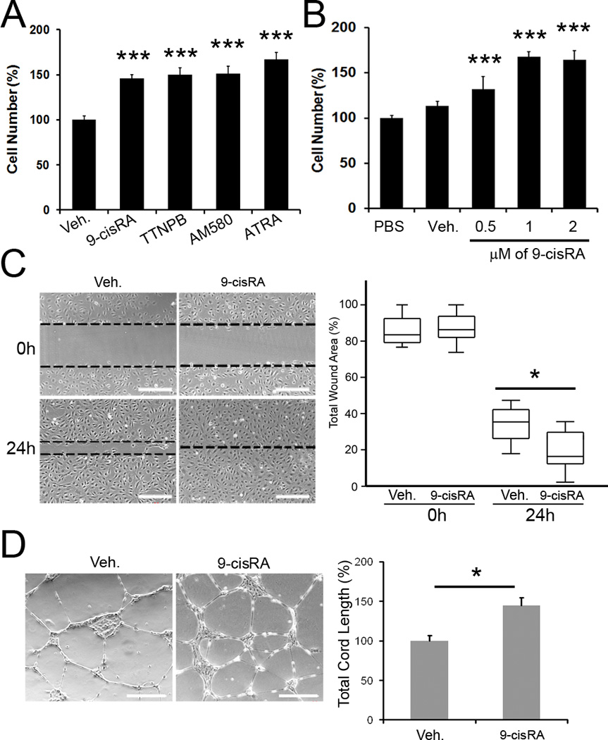
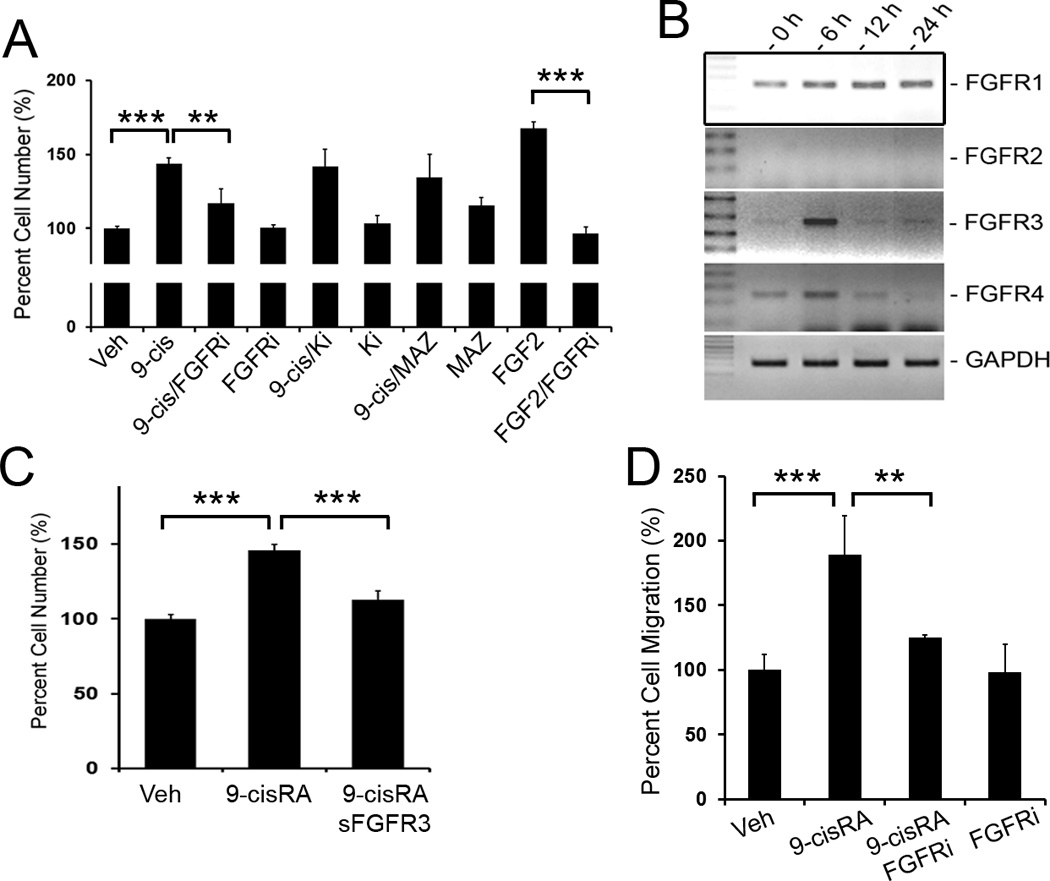
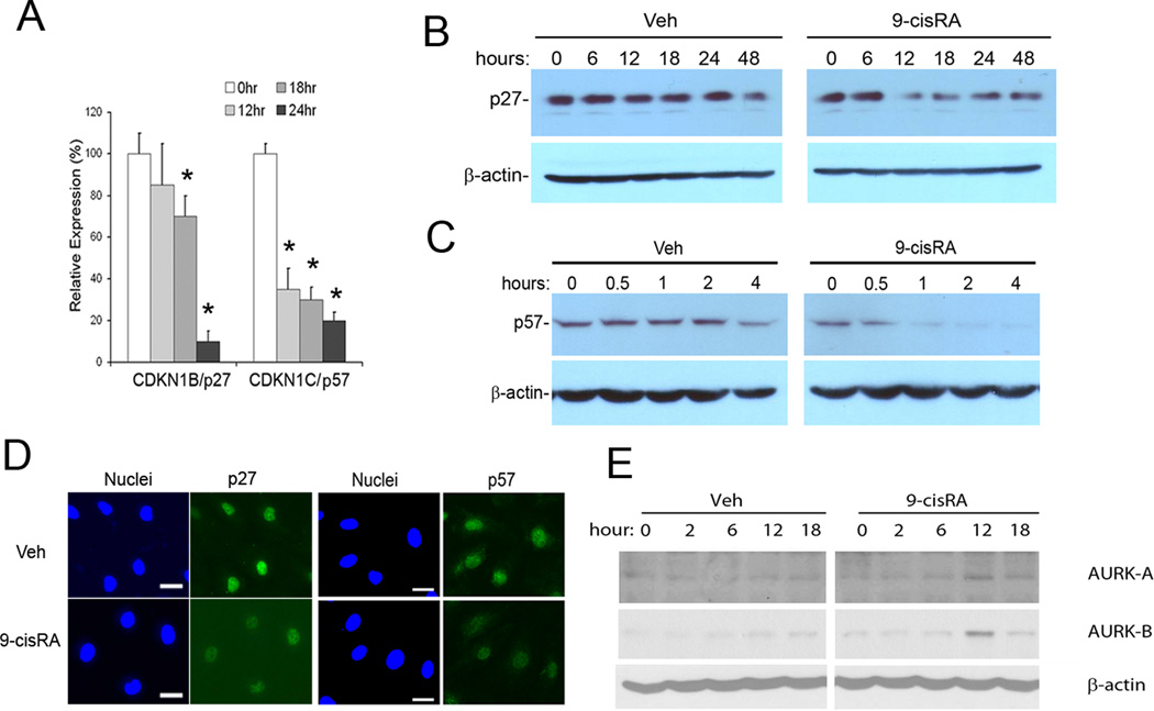
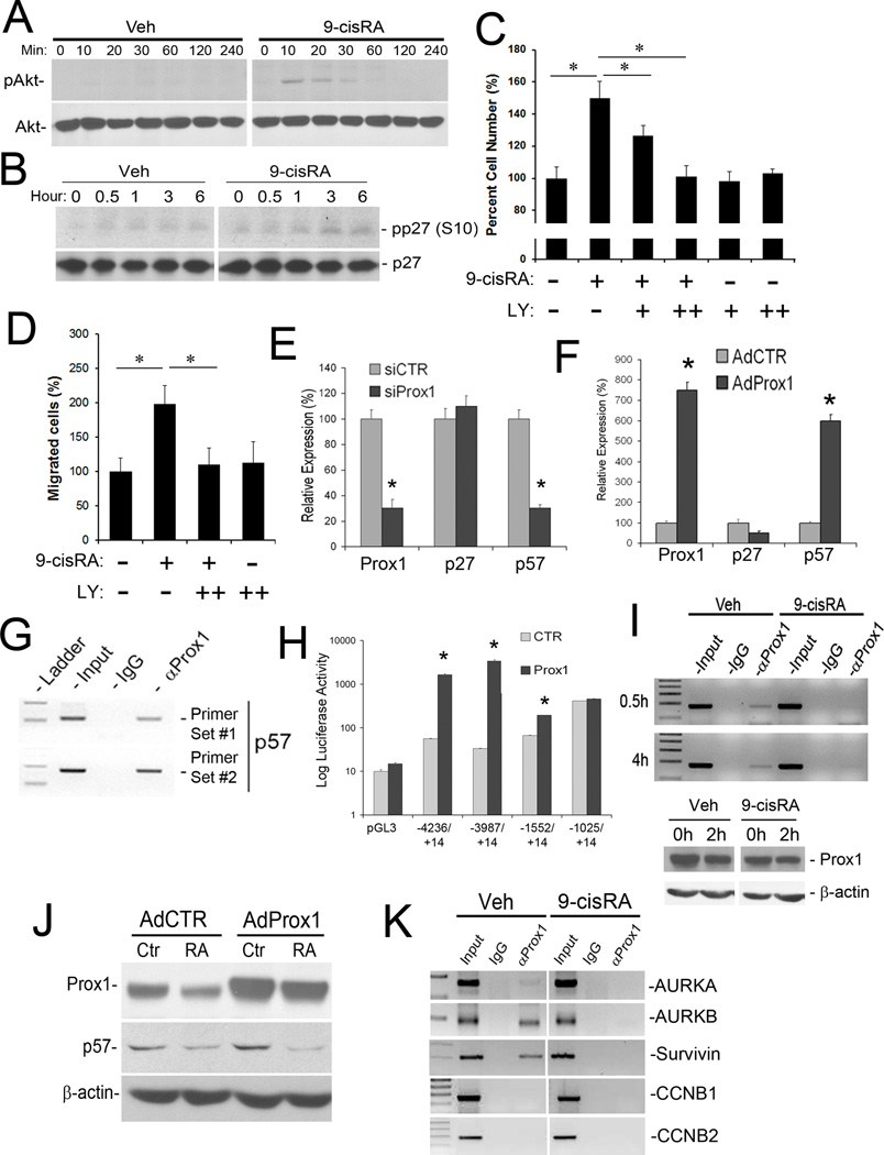
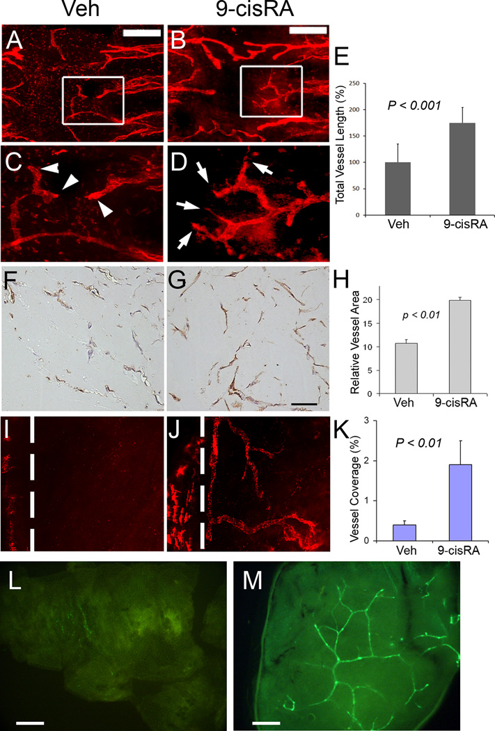
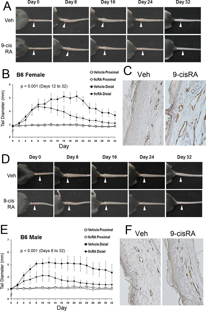
Methods and results: We report that RAs promote proliferation, migration, and tube formation of cultured lymphatic endothelial cells by activating fibroblast growth factor receptor signaling. Moreover, RAs control the expression of cell-cycle checkpoint regulators such as p27(Kip1), p57(Kip2), and the aurora kinases through both an Akt-mediated nongenomic action and a transcription-dependent genomic action that is mediated by Prox1, a master regulator of lymphatic development. Moreover, 9-cisRA was found to activate in vivo lymphangiogenesis in animals in mouse trachea, Matrigel plug, and cornea pocket assays. Finally, we demonstrate that 9-cisRA can provide a strong therapeutic efficacy in ameliorating experimental mouse tail lymphedema by enhancing lymphatic vessel regeneration.

Conclusion: These in vitro and animal studies demonstrate that 9-cisRA potently activates lymphangiogenesis and promotes lymphatic regeneration in an experimental lymphedema model, presenting it as a promising novel therapeutic agent to treat human lymphedema patients.


Conflict of interest statement

Conflict of Interest Disclosures: None

Figures

Figure 1
Figure 1
Retinoic acids activate proliferation, migration and tube formation of primary human LECs. (A) Activation of LEC-proliferation by various RA derivatives. Primary human LECs in a low serum media (1% FBS) were incubated with 1 µM of 9-cisRA, TTNPB, AM580, all-trans RA (ATRA) or vehicle (ethanol, 0.1%) alone. After 48 hours, the total cell number was estimated and displayed as a percent cell number against the vehicle alone (Veh)-treated control group. Bars represent the standard deviation (SD) of quadruplicates. Asterisks indicate p< 0.001 against the vehicle alone control. (B) Dose-dependent activation of LEC-proliferation in a low serum media (1% FBS) by 9-cisRA compared to phosphate-buffered saline (PBS) or vehicle alone (Veh) for 48 hours. Bars represent the standard deviation (SD) of quadruplicates. Asterisks indicate p< 0.001 against the vehicle alone control (Veh). (C) Effect of 9-cisRA on the migration of primary LECs. Monolayers of LECs were pre-treated with vehicle alone or vehicle containing 9-cisRA (1 µM) and scratched with a pipette tip to make a wound. After 24 hours, the total remaining wounded area was determined by image analysis and expressed as a percentage of the total wounded area in the box and whisker plot. Asterisk, p< 0.05. (D) Effect of 9-cisRA on tube formation of primary LECs. Cells were pre-treated with vehicle alone or vehicle with 9-cisRA (1 µM) for 24 hours and an equal number of cells was seeded onto the growth factor-depleted matrigel. After 24 hours, five representative images were taken and the total tube length per image was calculated and charted as a percentage of the vehicle-treated group. Asterisk, p< 0.05.
Figure 2
Figure 2
FGF pathway plays a key role in 9-cisRA-induced LEC-proliferation. (A) 9-cisRA-induced proliferation for 48 hours was significantly abrogated by a FGF-receptor inhibitor (FGFRi, PD173074, 50 nM) , but not by inhibitors for VEGFR-2 (Ki8751, 50 nM) or VEGFR-3 (MAZ51, 50 nM) . Three asterisks, p< 0.001; two asterisks, p < 0.01. (B) Expression of four FGF receptors (FGFR1 to FGFR4) and GAPDH (internal control) in LECs treated with 9-cisRA (1 µM) for 0, 6, 12 and 24 hours was determined by semi-quantitative RT-PCR. None of these genes was regulated by vehicle alone treatment (data not shown). (C) Pre-incubation of LECs with a soluble FGFR3 recombinant protein (1 µg/ml) reduced the 9-cisRA-promoted LEC proliferation. Three asterisks, p< 0.001. (D) Chemotactic migration assay using modified Boyden chambers (Fluoroblok™). Primary LECs were allowed to migrate toward serum-free media containing vehicle alone, 9-cisRA (1 µM), FGFR inhibitor (FGFRi, 50 nM) or 9-cisRA (1 µM)/FGFR inhibitor (FGFRi, 50 nM) for 3 hours. The migrated cells were quantified and expressed as a percentage against the vehicle control. Three asterisks, p< 0.001; two asterisks, p< 0.01.
Figure 3
Figure 3
Regulation of p27Kip1, p57Kip2 and aurora kinases by 9-cisRA in LECs. (A) qRT-PCR analyses show that the expression of CDKN1B/p27Kip1 and CDKN1C/p57Kip2 was significantly downregulated in 9-cisRA (1 µM) - treated LECs. Asterisk, p< 0.01 against 0 hour control; bars, the standard deviation (SD) of quadruplicates. Vehicle alone treatment did not alter the expression (data not shown). Western blot analyses also revealed 9-cisRA-mediated downregulation of the protein expression of p27Kip1 (B) and p57Kip2 (C). (D). Immunofluorescence staining assays demonstrated the downregulation of p27Kip1 and p57Kip2 after 18-hour of 9-cisRA (1 µM) treatment. Bars, 20 µm. (E) Protein expression of the aurora kinases A and B were transiently upregulated at 12 hours in LECs treated with 9-cisRA (1 µM).
Figure 4
Figure 4
Mechanism for 9-cisRA-mediated regulation of p27Kip1, p57Kip2 and the aurora kinases in LECs. (A) Non-genomic action of 9-cisRA on the rapid activation of Akt in LECs. The Akt protein is phosphorylated in LECs within 10 minutes of 9-cisRA (1 µM) treatment. (B) Phosphorylation of p27Kip1 at serine 10 (pp27(S10)) occurs from the first hour of 9-cisRA treatment of LECs. Expression of the whole p27Kip1 protein was also shown. (C) Essential role of the Akt-mediated non-genomic action of 9-cisRA in LEC proliferation. LECs were pretreated with the PI3K signal inhibitor LY294002 (LY 50 nM (+), 100 nM (++)) for 30 minutes before 9-cisRA treatment (1 µM) and allowed to grow for 48 hours before determination of the relative cell numbers. Note that the inhibition of the PI3K/Akt signal blocked 9-cisRA-induced LEC proliferation. (D) Similarly, LECs were pretreated with LY294002 (LY, 100 nM (++)) for 30 minutes before 9-cisRA treatment (1 µM) and allowed to migrate in the modified Boyden chamber for 3 hours before measurement of the relative number of migrated cells. Note the essential role of the Akt-mediated non-genomic action of 9-cisRA in LEC migration. (E) qRT-PCR assays showed that knockdown of Prox1 in LECs resulted in downregulation of p57Kip2, but not p27Kip1. (F) Adenoviral overexpression of Prox1 (AdProx1) in HUVECs for 48 hours strongly induced p57Kip2, but not p27Kip1, as determined by qRT-PCR. AdCTR, control adenovirus. (G) ChIP assays for Prox1 in LECs performed against the p57Kip2 promoter using two independent PCR primer sets showed a physical association of the Prox1 protein with the p57Kip2 promoter. (H) Luciferase-reporter assays measuring the response of the different length proximal promoters of p57Kip2 to Prox1-mediated activation. The reporter constructs were named by the position of its 5’ and 3’ ends of the promoter fragments relative to the transcriptional start site (+1) of p57Kip2. (I) Prox1 ChIP was performed against the p57Kip2 promoter from LECs treated with vehicle (Veh) or 9-cisRA (1 µM) for 0.5 or 4 hours. The expression level of Prox1 protein was also determined in LECs treated with 9-cisRA for 2 hours. (J) Western blot assay showing that overexpression of Prox1 in the presence of 9-cisRA did not induce the expression of p57Kip2. (K) Prox1 ChIP revealed that, upon treatment of LECs with 9-cisRA (1 µM, 4 hours), Prox1 dissociated from the promoters of the aurora kinase kinases A (AURKA) and B (AURKB) and Survivin. Prox1 did not bind to the promoters of CCNB1 and CCNB2. For panels C, D, E, F and H, data are shown as the mean ± standard deviation (SD) and asterisks present p< 0.05.
Figure 5
Figure 5
Promotion of in vivo lymphangiogenesis by 9-cisRA. (A–E) Mouse trachea assay: Lymphangiogenesis was activated in 9-cisRA-treated trachea (B, D) (5 mice) compared to vehicle-treated trachea (A, C) (5 mice). Low magnification images (A, B) of the tracheal mucosa are shown (Bars, 400 µm). Enlarged images of the boxed areas in panels A and B show that the lymphatic vessels in the vehicle-treated trachea are round-ended (arrowheads in C), whereas the lymphatics in the 9-cisRA-treated trachea have jagged ends (arrows in D), indicating active lymphatic sprouting. (E) Total length of the lymphatic vessels was measured on the luminal side of the trachealis muscle, which is largely devoid of lymphatic vessels. (F–H) Mouse subcutaneous matrigel plug assay: Matrigel premixed with vehicle alone (F) or 9-cisRA (10 µM) (G) was subcutaneously injected (two matrigel plugs per mouse, 5 mice each group) and harvested after two weeks for immunohistochemistry (IHC) analyses using an anti-podoplanin antibody. The relative lymphatic vessel area was determined in the matrigel using the NIH ImageJ software (H). Bar in (G), 50 µm. (I–K) 9-cisRA stimulates lymphangiogenesis in the mouse cornea. Representative LYVE-1 (red) immunofluorescent images are shown to demonstrate lymphatic vessel ingrowth into the cornea implanted with vehicle pellet (I) or 9-cisRA pellet (J) after 14 days. Dotted line: demarcation between the cornea and conjunctiva. Original Magnification: X100. (K) A significant difference was observed in the lymphatic coverage area between the vehicle (10 mice) and 9-cisRA (13 mice) groups. Error bars represent S.E.M. (L, M) Subcutaneous matrigel plug assay using the previously reported lymphatic-specific GFP mice to visualize the newly formed lymphatic networks: Matrigel premixed with vehicle alone (L) or vehicle containing 9-cisRA (10 µM) (M) was injected into the skin of the lymphatic-specific GFP mice (two matrigel plugs per mouse, 5 mice each group) and isolated after two weeks. Lymphatic vessels within the matrigels were directly visualized under a fluorescent stereoscope.
Figure 6
Figure 6
Therapeutic effect of 9-cisRA on secondary lymphedema and lymphatic vessel regeneration. Experimental lymphedema was surgically induced in the tail of C57BL/6J mice and vehicle alone (ethanol/seed oil) or vehicle with 9-cisRA (0.8 mg/kg) was i.p. injected daily from day 2. Images of secondary lymphedema development in female (A–C) and male (D–F) mice treated with vehicle (Veh) or 9-cisRA (9cRA) for 32 days. The diameter of the tail on the distal (white arrowheads) and proximal sides of the wounds was measured every other day and charted for the female (B) and male (E) mice. Six females and 5 males were used for the vehicle-treated group and 7 females and 6 males were for the 9-cisRA-treated group. Cross, p< 0.05; asterisk, p< 0.01. (C, F) After 32 days, prominent regeneration of LYVE-1-positive lymphatic vessels was detected in the 9-cisRA-treated mice, compared to the vehicle-treated group (Veh), in both genders. For statistical analyses, a mixed linear model with the autoregressive covariance structure overtime was used to compare tail diameters over time by treatment and distal side of the wounds. Statistically significant difference (p< 0.001) was observed for the distal side of the wounds in females from days 12 to 32 and in males from days 6 to 32. No statistical difference was found for the proximal side of the wounds between the two groups in both genders. Similar data was obtained from an experiment using BALB/c mice (Supplemental Figure 1).

Comment in

Similar articles

Cited by

References

    1. Wigle JT, Oliver G. Prox1 function is required for the development of the murine lymphatic system. Cell. 1999;98:769–778. - PubMed
    1. Wigle JT, Harvey N, Detmar M, Lagutina I, Grosveld G, Gunn MD, Jackson DG, Oliver G. An essential role for prox1 in the induction of the lymphatic endothelial cell phenotype. EMBO J. 2002;21:1505–1513. - PMC - PubMed
    1. Lahteenvuo M, Honkonen K, Tervala T, Tammela T, Suominen E, Lahteenvuo J, Kholova I, Alitalo K, Yla-Herttuala S, Saaristo A. Growth factor therapy and autologous lymph node transfer in lymphedema. Circulation. 2011;123:613–620. - PubMed
    1. Tammela T, Saaristo A, Holopainen T, Lyytikka J, Kotronen A, Pitkonen M, Abo-Ramadan U, Yla-Herttuala S, Petrova TV, Alitalo K. Therapeutic differentiation and maturation of lymphatic vessels after lymph node dissection and transplantation. Nat Med. 2007;13:1458–1466. - PubMed
    1. Szanto A, Narkar V, Shen Q, Uray IP, Davies PJ, Nagy L. Retinoid x receptors: X-ploring their (patho)physiological functions. Cell Death Differ. 2004;11 Suppl 2:S126–S143. - PubMed

Publication types

MeSH terms