. 2012 Mar 15;18(6):1641-54.
doi: 10.1158/1078-0432.CCR-11-2324. Epub 2012 Jan 24.

Combined MEK and VEGFR inhibition in orthotopic human lung cancer models results in enhanced inhibition of tumor angiogenesis, growth, and metastasis

Affiliations

Combined MEK and VEGFR inhibition in orthotopic human lung cancer models results in enhanced inhibition of tumor angiogenesis, growth, and metastasis

Osamu Takahashi et al. Clin Cancer Res. .

Abstract

Purpose: Ras/Raf/mitogen-activated protein-extracellular signal-regulated kinase (ERK) kinase (MEK)/ERK signaling is critical for tumor cell proliferation and survival. Selumetinib is a potent, selective, and orally available MEK1/2 inhibitor. In this study, we evaluated the therapeutic efficacy of selumetinib alone or with cediranib, an orally available potent inhibitor of all three VEGF receptor (VEGFR) tyrosine kinases, in murine orthotopic non-small cell lung carcinoma (NSCLC) models.

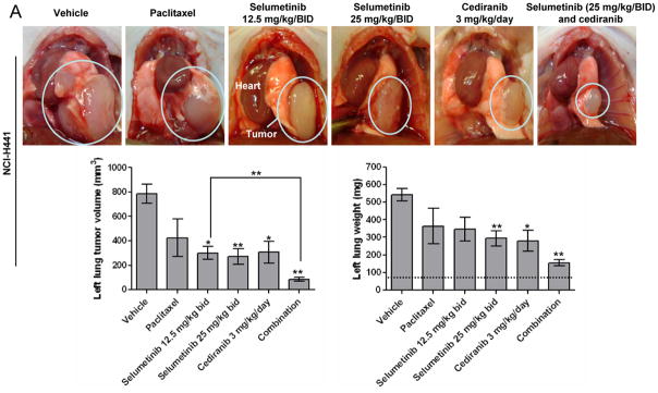
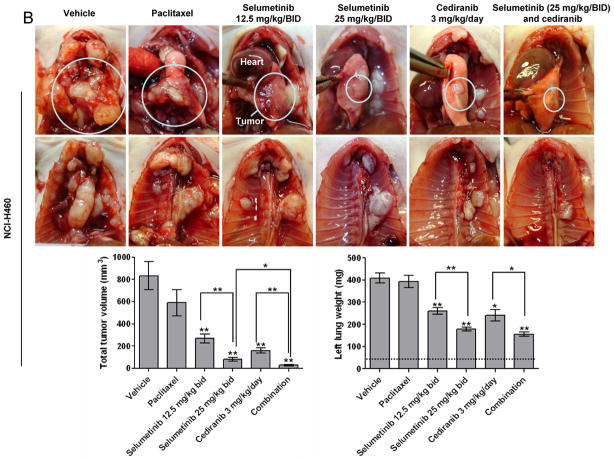
Experimental design: NCI-H441 or NCI-H460 KRAS-mutant human NSCLC cells were injected into the lungs of mice. Mice were randomly assigned to treatment with selumetinib, cediranib, paclitaxel, selumetinib plus cediranib, or control. When controls became moribund, all animals were sacrificed and assessed for lung tumor burden and locoregional metastasis. Lung tumors and adjacent normal tissues were subjected to immunohistochemical analyses.

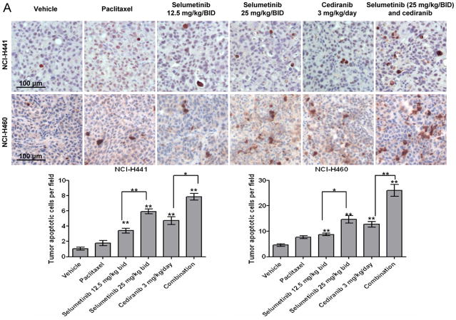
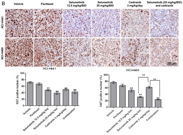
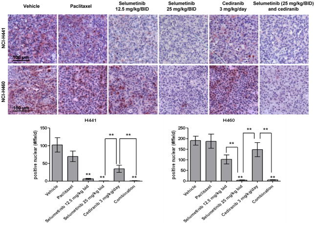
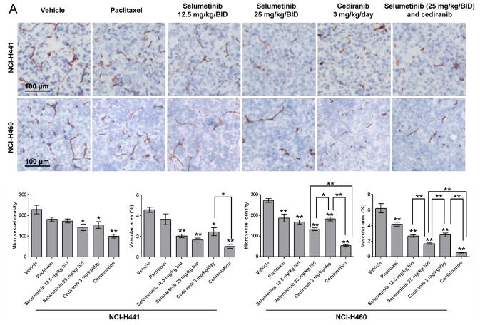
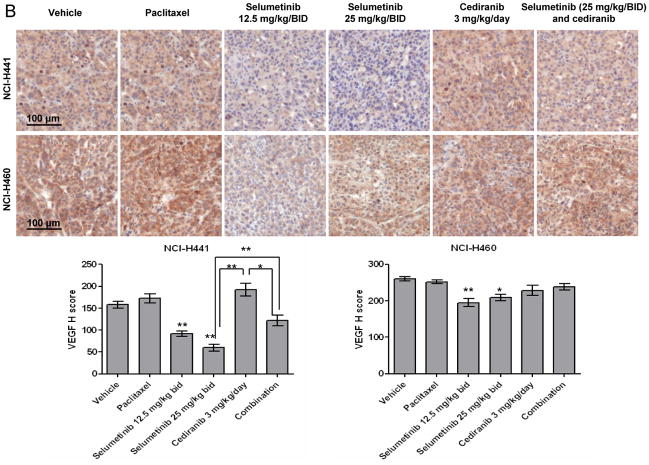
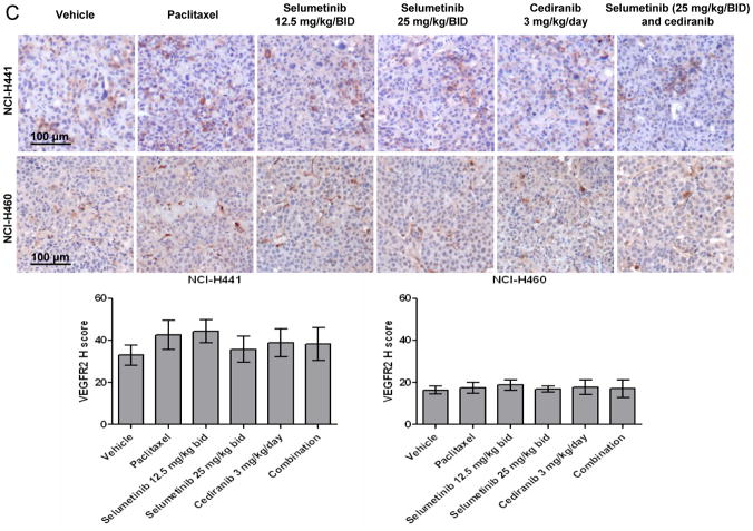
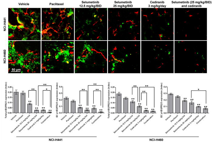
Results: Selumetinib inhibited lung tumor growth and, particularly at higher dose, reduced locoregional metastasis, as did cediranib. Combining selumetinib and cediranib markedly enhanced their antitumor effects, with near complete suppression of metastasis. Immunohistochemistry of tumor tissues revealed that selumetinib alone or with cediranib reduced ERK phosphorylation, angiogenesis, and tumor cell proliferation and increased apoptosis. The antiangiogenic and apoptotic effects were substantially enhanced when the agents were combined. Selumetinib also inhibited lung tumor VEGF production and VEGFR signaling.

Conclusions: In this study, we evaluated therapy directed against MEK combined with antiangiogenic therapy in distinct orthotopic NSCLC models. MEK inhibition resulted in potent antiangiogenic effects with decreased VEGF expression and signaling. Combining selumetinib with cediranib enhanced their antitumor and antiangiogenic effects. We conclude that combining selumetinib and cediranib represents a promising strategy for the treatment of NSCLC.


Figures

Figure 1
Figure 1
Anti-tumor effects of selumetinib, cediranib, and paclitaxel in orthotopic models of human NSCLC in mice. *p < 0.007 or **p < 0.001 versus vehicle control or between groups as indicated (Kruskal-Wallis followed by Mann-Whitney U tests). A. Primary tumors in the left lungs of representative mice from each treatment group after implantation of NCI-H441 human lung adenocarcinoma cells (circled), with mean primary tumor lung volumes and left lung weights. Bars indicate standard error of the mean. B. Primary tumors in the left lungs (circled) and chest walls of representative mice from each treatment group after implantation of NCI-H460 human lung large cell cancer cells, with mean total tumor volumes (primary lung tumor + chest wall tumors) and left lung weights. Bars indicate standard error of the mean. The dotted horizontal line indicates the normal left lung weight.
Figure 1
Figure 1
Anti-tumor effects of selumetinib, cediranib, and paclitaxel in orthotopic models of human NSCLC in mice. *p < 0.007 or **p < 0.001 versus vehicle control or between groups as indicated (Kruskal-Wallis followed by Mann-Whitney U tests). A. Primary tumors in the left lungs of representative mice from each treatment group after implantation of NCI-H441 human lung adenocarcinoma cells (circled), with mean primary tumor lung volumes and left lung weights. Bars indicate standard error of the mean. B. Primary tumors in the left lungs (circled) and chest walls of representative mice from each treatment group after implantation of NCI-H460 human lung large cell cancer cells, with mean total tumor volumes (primary lung tumor + chest wall tumors) and left lung weights. Bars indicate standard error of the mean. The dotted horizontal line indicates the normal left lung weight.
Figure 2
Figure 2
Apoptotic and anti-proliferative effects of selumetinib, cediranib, and paclitaxel in orthotopic models of human NSCLC in mice. Lung tumors were collected 2 hours after the last dose of selumetinib, 20 hours after the last dose of cediranib, and 6 days after the last dose of paclitaxel. Quantitative values were determined in four random fields for each tumor. Data are presented as means ± SEM for 9–13 samples per group. The scale bar indicates 100 μm. *p < 0.007, **p < 0.001 versus vehicle control or between groups as indicated (Kruskal-Wallis followed by Mann-Whitney U tests). A. Immunohistochemical analysis of cleaved caspase 3 in tumors from mice implanted with NCI-H441 or NCI-H460 cells. Representative cleaved caspase-3 staining (brown staining in positive cells) in lung tumors viewed at x200 magnification. Tumor cell apoptosis (number of cleaved caspase-3 positive cells/field) is shown. B. Immunohistochemical analysis of Ki67 staining in tumors from mice implanted with NCI-H441 or NCI-H460 cells. Representative Ki67 staining of lung tumors (brown) viewed at x100 magnification, 60% center field. Tumor proliferation (percentage of Ki67-positive cells/total tumor cells) is shown.
Figure 2
Figure 2
Apoptotic and anti-proliferative effects of selumetinib, cediranib, and paclitaxel in orthotopic models of human NSCLC in mice. Lung tumors were collected 2 hours after the last dose of selumetinib, 20 hours after the last dose of cediranib, and 6 days after the last dose of paclitaxel. Quantitative values were determined in four random fields for each tumor. Data are presented as means ± SEM for 9–13 samples per group. The scale bar indicates 100 μm. *p < 0.007, **p < 0.001 versus vehicle control or between groups as indicated (Kruskal-Wallis followed by Mann-Whitney U tests). A. Immunohistochemical analysis of cleaved caspase 3 in tumors from mice implanted with NCI-H441 or NCI-H460 cells. Representative cleaved caspase-3 staining (brown staining in positive cells) in lung tumors viewed at x200 magnification. Tumor cell apoptosis (number of cleaved caspase-3 positive cells/field) is shown. B. Immunohistochemical analysis of Ki67 staining in tumors from mice implanted with NCI-H441 or NCI-H460 cells. Representative Ki67 staining of lung tumors (brown) viewed at x100 magnification, 60% center field. Tumor proliferation (percentage of Ki67-positive cells/total tumor cells) is shown.
Figure 3
Figure 3
Effects of selumetinib, cediranib, and paclitaxel upon ERK signaling in orthotopic models of human NSCLC in mice. Representative pERK staining of lung tumors (purple) and quantified pERK expression (number of pERK positive cells/field). All stains are viewed at x100 magnification, 60% center field. Lung tumors were collected 2 hours after the last dose of selumetinib, 20 hours after the last dose of cediranib, and 6 days after the last dose of paclitaxel. Quantitative values were determined in four random fields for each tumor. Data are presented as means ± SEM for 9–13 samples per group. The scale bar indicates 100 μm. *p < 0.007, **p < 0.001 versus vehicle control or between groups as indicated (Kruskal-Wallis followed by Mann-Whitney U tests).
Figure 4
Figure 4
Antiangiogenic effects of selumetinib, cediranib, and paclitaxel in orthotopic models of human NSCLC in mice as assessed by immunohistochemical analysis of CD31. Representative CD31 staining of lung tumors (brown) viewed at x100 magnification, 60% center field, and microvessel density (number of CD31-positive objects/field) and vascular area (area of CD31-positive objects/field area × 100%) are shown. Lung tumors were collected 2 hours after the last dose of selumetinib, 20 hours after the last dose of cediranib, and 6 days after the last dose of paclitaxel. Quantitative values were determined in four random fields for each tumor. Data are presented as means ± SEM for 9–13 samples per group. The scale bar indicates 100 μm. *p < 0.007, **p < 0.001 versus vehicle control or between groups as indicated (Kruskal-Wallis followed by Mann-Whitney U tests).
Figure 5
Figure 5
Effects of selumetinib, cediranib, and paclitaxel upon VEGF and VEFGR expression in orthotopic models of human NSCLC in mice. Lung tumors were collected 2 hours after the last dose of selumetinib, 20 hours after the last dose of cediranib, and 6 days after the last dose of paclitaxel. Quantitative values were determined in four random fields for each tumor. Data are presented as means ± SEM for 9–13 samples per group. The scale bar indicates 100 μm. *p < 0.007, **p < 0.001 versus vehicle control or between groups as indicated (Kruskal-Wallis followed by Mann-Whitney U tests). A. Immunohistochemical analysis of VEGF in lung tumors from mice implanted with either NCI-H441 or NCI-H460 cells. Representative VEGF staining (brown cytoplasmic staining) in lung tumors and H scores for VEGF expression are shown. B. Immunohistochemical analysis of VEGR2 in lung tumors from mice implanted with either NCI-H441 or NCI-H460 cells. Representative VEGFR2 staining (brown cytoplasmic staining) in lung tumors and H scores for VEGFR2 expression are shown.
Figure 5
Figure 5
Effects of selumetinib, cediranib, and paclitaxel upon VEGF and VEFGR expression in orthotopic models of human NSCLC in mice. Lung tumors were collected 2 hours after the last dose of selumetinib, 20 hours after the last dose of cediranib, and 6 days after the last dose of paclitaxel. Quantitative values were determined in four random fields for each tumor. Data are presented as means ± SEM for 9–13 samples per group. The scale bar indicates 100 μm. *p < 0.007, **p < 0.001 versus vehicle control or between groups as indicated (Kruskal-Wallis followed by Mann-Whitney U tests). A. Immunohistochemical analysis of VEGF in lung tumors from mice implanted with either NCI-H441 or NCI-H460 cells. Representative VEGF staining (brown cytoplasmic staining) in lung tumors and H scores for VEGF expression are shown. B. Immunohistochemical analysis of VEGR2 in lung tumors from mice implanted with either NCI-H441 or NCI-H460 cells. Representative VEGFR2 staining (brown cytoplasmic staining) in lung tumors and H scores for VEGFR2 expression are shown.
Figure 6
Figure 6
Effects of selumetinib, cediranib, and paclitaxel upon VEFGR signaling in orthotopic models of human NSCLC in mice. CD31/pVEGFR2/3 dual fluorescent staining in tumors from mice implanted with either NCI-H441 or NCI-H460 cells was completed. Representative co-localized CD31/pVEGFR2/3 staining of lung tumors viewed at x200 magnification are shown. Fluorescent red indicates CD31-positive endothelial cells; fluorescent green, total pVEGFR2/3-positive cells; fluorescent yellow, pVEGFR2/3-positive endothelial cells. Quantification of total activated VEGFR2/3 expression is presented as an index of green area to blue area. Activated VEGFR2/3 expression in endothelial cells is presented as an index of yellow area to red area. All quantification data are presented as means ± SEM for 9–13 samples per group. The scale bar indicates 50 μm. *p < 0.007 or **p < 0.001 versus vehicle control or between groups as indicated (Kruskal-Wallis followed by Mann-Whitney U tests).

Similar articles

Cited by

References

    1. Jemal A, Siegel R, Xu J, Ward E. Cancer statistics, 2010. CA Cancer J Clin. 2010;60:277–300. - PubMed
    1. Parkin DM, Bray F, Ferlay J, Pisani P. Global Cancer Statistics, 2002. CA Cancer J Clin. 2005;55:74–108. - PubMed
    1. Pao W, Girard N. New driver mutations in non-small-cell lung cancer. Lancet Oncol. 2011;12:175–80. - PubMed
    1. Sandler A, Gray R, Perry MC, Brahmer J, Schiller JH, Dowlati A, et al. Paclitaxel-carboplatin alone or with bevacizumab for non-small-cell lung cancer. N Engl J Med. 2006;355:2542–50. - PubMed
    1. Reck M, von Pawel J, Zatloukal P, Ramlau R, Gorbounova V, Hirsh V, et al. Phase III Trial of Cisplatin Plus Gemcitabine With Either Placebo or Bevacizumab As First-Line Therapy for Nonsquamous Non-Small-Cell Lung Cancer: AVAiL. J Clin Oncol. 2009 - PubMed

Publication types

MeSH terms