

Insulin-like growth factor 1 alleviates high-fat diet-induced myocardial contractile dysfunction: role of insulin signaling and mitochondrial function
- PMID: 22275536
- PMCID: PMC3288378
- DOI: 10.1161/HYPERTENSIONAHA.111.181867
Insulin-like growth factor 1 alleviates high-fat diet-induced myocardial contractile dysfunction: role of insulin signaling and mitochondrial function
Erratum in
- Hypertension. 2014 Apr;63(4):e92
Retraction in
-
Retraction of: Insulin-Like Growth Factor 1 Alleviates High-Fat Diet-Induced Myocardial Contractile Dysfunction: Role of Insulin Signaling and Mitochondrial Function.Hypertension. 2022 Sep;79(9):e115. doi: 10.1161/HYP.0000000000000219. Epub 2022 Jul 22. Hypertension. 2022. PMID: 35866416 Free PMC article. No abstract available.
Abstract
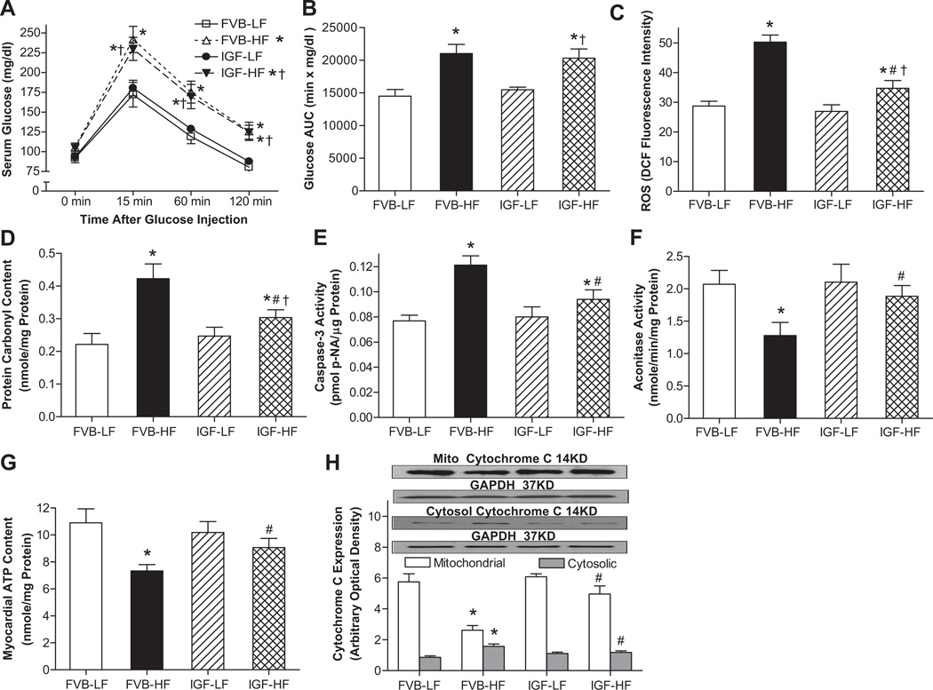
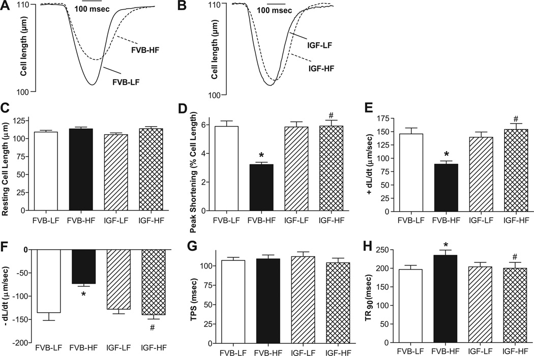
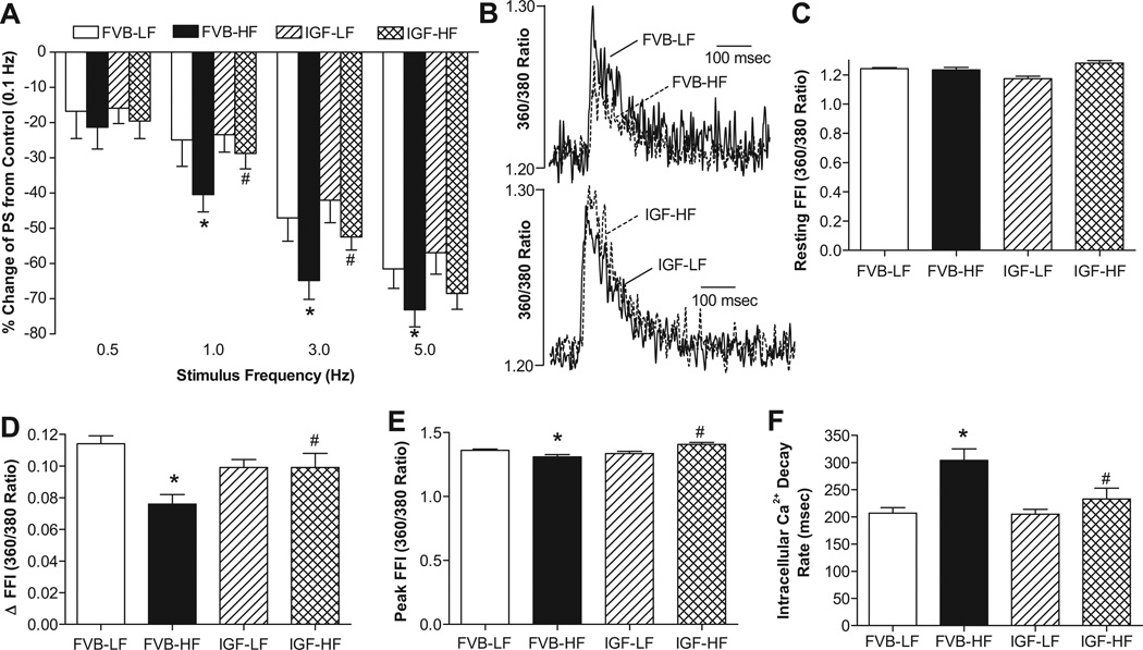
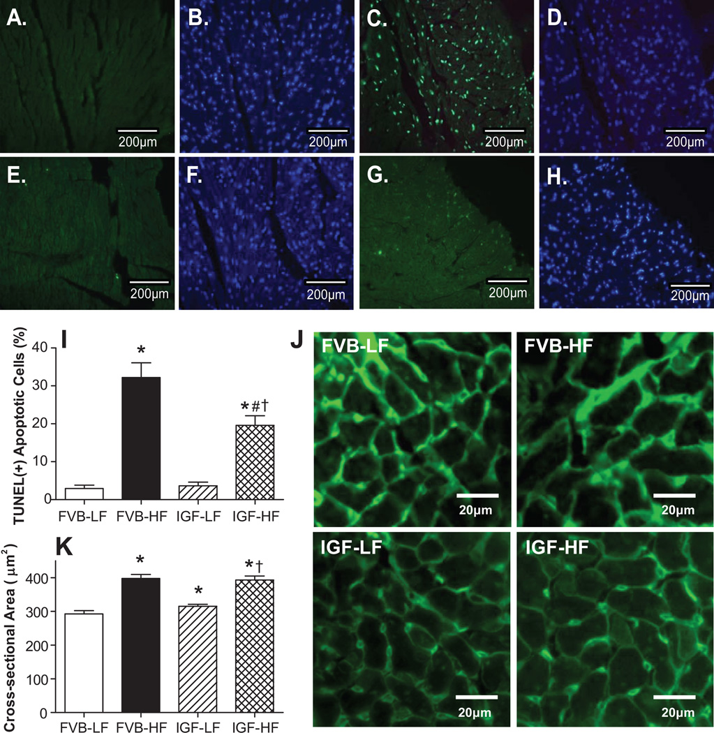
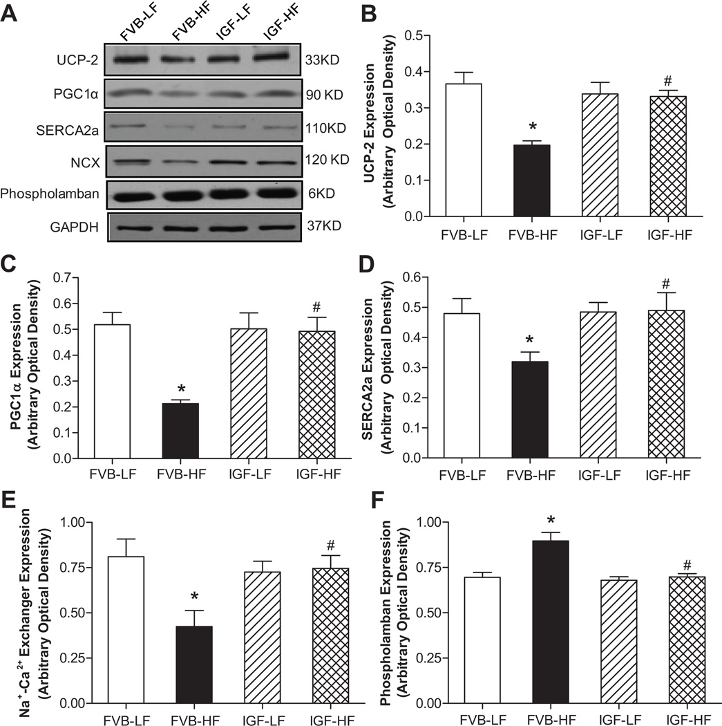
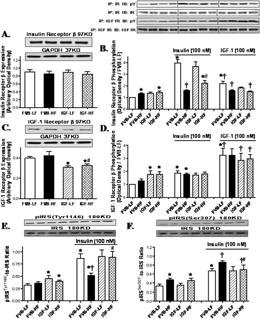
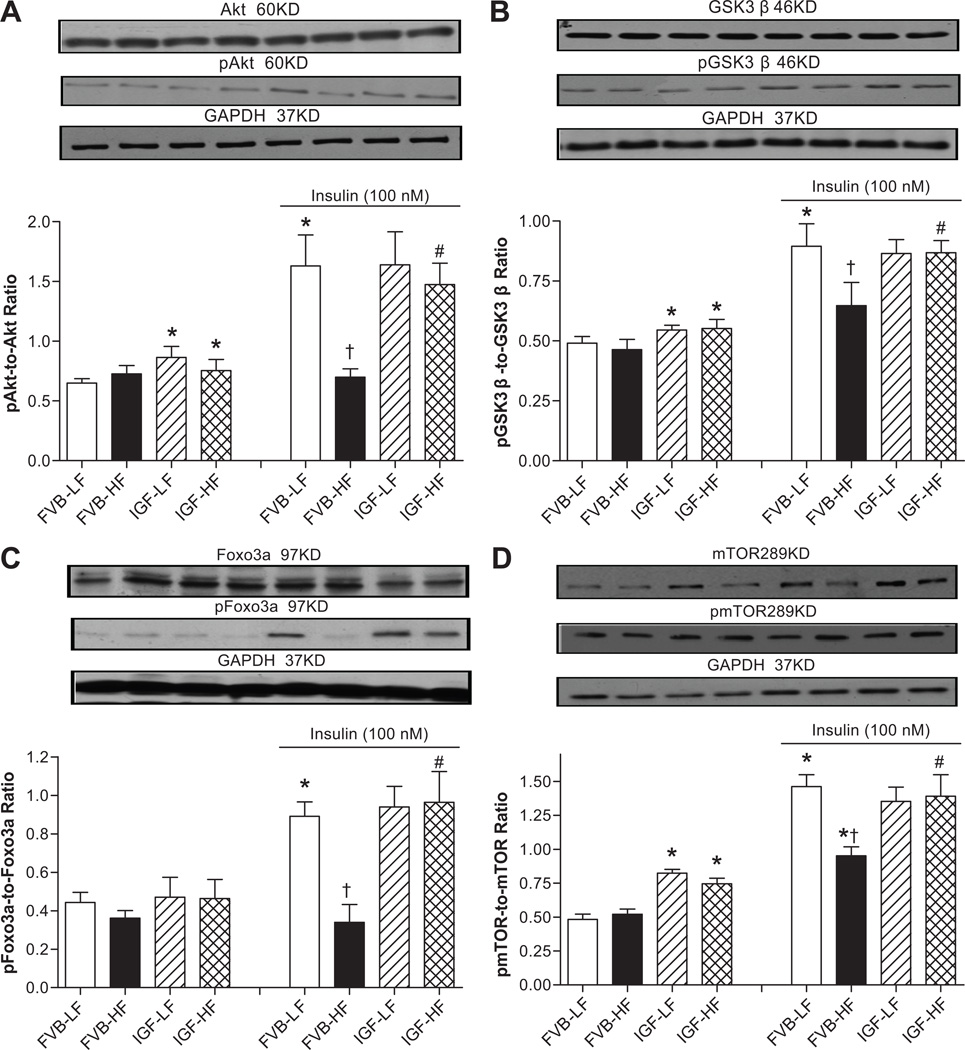
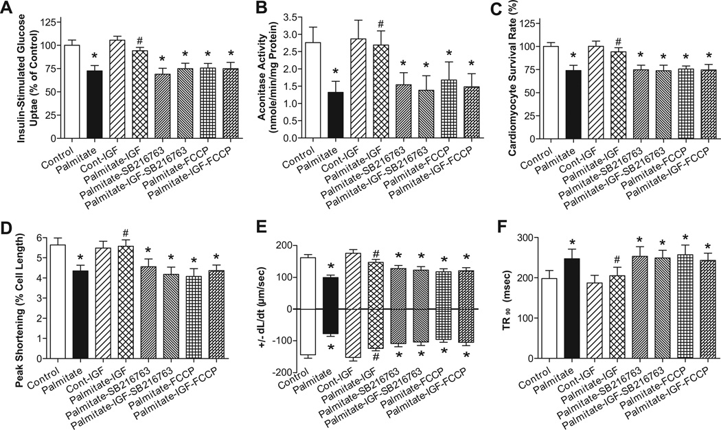
Obesity is often associated with reduced plasma insulin-like growth factor 1 (IGF-1) levels, oxidative stress, mitochondrial damage, and cardiac dysfunction. This study was designed to evaluate the impact of IGF-1 on high-fat diet-induced oxidative, myocardial, geometric, and mitochondrial responses. FVB and cardiomyocyte-specific IGF-1 overexpression transgenic mice were fed a low- (10%) or high-fat (45%) diet to induce obesity. High-fat diet feeding led to glucose intolerance, elevated plasma levels of leptin, interleukin 6, insulin, and triglyceride, as well as reduced circulating IGF-1 levels. Echocardiography revealed reduced fractional shortening, increased end-systolic and end-diastolic diameter, increased wall thickness, and cardiac hypertrophy in high-fat-fed FVB mice. High-fat diet promoted reactive oxygen species generation, apoptosis, protein and mitochondrial damage, reduced ATP content, cardiomyocyte cross-sectional area, contractile and intracellular Ca(2+) dysregulation (including depressed peak shortening and maximal velocity of shortening/relengthening), prolonged duration of relengthening, and dampened intracellular Ca(2+) rise and clearance. Western blot analysis revealed disrupted phosphorylation of insulin receptor and postreceptor signaling molecules insulin receptor substrate 1 (tyrosine/serine phosphorylation), Akt, glycogen synthase kinase 3β, forkhead transcriptional factors, and mammalian target of rapamycin, as well as downregulated expression of mitochondrial proteins peroxisome proliferator-activated receptor-γ coactivator 1α and uncoupling protein 2. Intriguingly, IGF-1 mitigated high-fat-diet feeding-induced alterations in reactive oxygen species, protein and mitochondrial damage, ATP content, apoptosis, myocardial contraction, intracellular Ca(2+) handling, and insulin signaling but not whole body glucose intolerance and cardiac hypertrophy. Exogenous IGF-1 treatment also alleviated high-fat diet-induced cardiac dysfunction. Our data revealed that IGF-1 alleviates high-fat diet-induced cardiac dysfunction despite persistent cardiac remodeling, possibly because of preserved cell survival, mitochondrial function, and insulin signaling.
Figures
Comment in
-
Heart smart insulin-like growth factor 1.Hypertension. 2012 Mar;59(3):550-1. doi: 10.1161/HYPERTENSIONAHA.111.188441. Epub 2012 Jan 23. Hypertension. 2012. PMID: 22275529 No abstract available.
Similar articles
-
Metallothionein prevents high-fat diet induced cardiac contractile dysfunction: role of peroxisome proliferator activated receptor gamma coactivator 1alpha and mitochondrial biogenesis.Diabetes. 2007 Sep;56(9):2201-12. doi: 10.2337/db06-1596. Epub 2007 Jun 15. Diabetes. 2007. PMID: 17575086
-
Mitochondrial aldehyde dehydrogenase (ALDH2) protects against streptozotocin-induced diabetic cardiomyopathy: role of GSK3β and mitochondrial function.BMC Med. 2012 Apr 23;10:40. doi: 10.1186/1741-7015-10-40. BMC Med. 2012. Retraction in: BMC Med. 2022 Jul 8;20(1):248. doi: 10.1186/s12916-022-02455-5. PMID: 22524197 Free PMC article. Retracted.
-
Tauroursodeoxycholic acid mitigates high fat diet-induced cardiomyocyte contractile and intracellular Ca2+ anomalies.PLoS One. 2013 May 7;8(5):e63615. doi: 10.1371/journal.pone.0063615. Print 2013. PLoS One. 2013. PMID: 23667647 Free PMC article.
-
Moderate ethanol administration accentuates cardiomyocyte contractile dysfunction and mitochondrial injury in high fat diet-induced obesity.Toxicol Lett. 2015 Mar 18;233(3):267-77. doi: 10.1016/j.toxlet.2014.12.018. Epub 2014 Dec 27. Toxicol Lett. 2015. PMID: 25549548
-
MitomiRs Keep the Heart Beating.Adv Exp Med Biol. 2017;982:431-450. doi: 10.1007/978-3-319-55330-6_23. Adv Exp Med Biol. 2017. PMID: 28551801 Review.
Cited by
-
Nutritional models of foetal programming and nutrigenomic and epigenomic dysregulations of fatty acid metabolism in the liver and heart.Pflugers Arch. 2014 May;466(5):833-50. doi: 10.1007/s00424-013-1339-4. Epub 2013 Sep 3. Pflugers Arch. 2014. PMID: 23999818 Review.
-
A novel function of CREG in metabolic disorders.Med Rev (2021). 2022 Jan 11;1(1):18-22. doi: 10.1515/mr-2021-0031. eCollection 2021 Oct. Med Rev (2021). 2022. PMID: 37724076 Free PMC article. Review.
-
Mitochondrial aldehyde dehydrogenase obliterates insulin resistance-induced cardiac dysfunction through deacetylation of PGC-1α.Oncotarget. 2016 Nov 22;7(47):76398-76414. doi: 10.18632/oncotarget.11977. Oncotarget. 2016. PMID: 27634872 Free PMC article.
-
Obesity and insulin resistance induce early development of diastolic dysfunction in young female mice fed a Western diet.Endocrinology. 2013 Oct;154(10):3632-42. doi: 10.1210/en.2013-1256. Epub 2013 Jul 24. Endocrinology. 2013. PMID: 23885014 Free PMC article.
-
Insulin Resistance Is Cheerfully Hitched with Hypertension.Life (Basel). 2022 Apr 10;12(4):564. doi: 10.3390/life12040564. Life (Basel). 2022. PMID: 35455055 Free PMC article. Review.
References
-
- Eckel RH, Barouch WW, Ershow AG. Report of the National Heart, Lung, and Blood Institute-National Institute of Diabetes and Digestive and Kidney Diseases Working Group on the pathophysiology of obesity-associated cardiovascular disease. Circulation. 2002;105:2923–2928. - PubMed
-
- Li SY, Yang X, Ceylan-Isik AF, Du M, Sreejayan N, Ren J. Cardiac contractile dysfunction in Lep/Lep obesity is accompanied by NADPH oxidase activation, oxidative modification of sarco(endo)plasmic reticulum Ca2+-ATPase and myosin heavy chain isozyme switch. Diabetologia. 2006;49:1434–1446. - PubMed
-
- de Divitiis O, Fazio S, Petitto M, Maddalena G, Contaldo F, Mancini M. Obesity and cardiac function. Circulation. 1981;64:477–482. - PubMed
-
- Zhang Y, Ren J. Role of cardiac steatosis and lipotoxicity in obesity cardiomyopathy. Hypertension. 2011;57:148–150. - PubMed
Publication types
MeSH terms
Substances
Grants and funding
LinkOut - more resources
Full Text Sources
Miscellaneous
