

doi: 10.1080/08998280.2012.11928791.
Papillary tumor of the pineal region
Affiliations
- PMID: 22275792
- PMCID: PMC3246862
- DOI: 10.1080/08998280.2012.11928791
Item in Clipboard
Papillary tumor of the pineal region
Proc (Bayl Univ Med Cent).
2012 Jan.
Abstract
Presented is a patient with papillary tumor of the pineal region (PTPR), an uncommon and recently recognized neoplasm. As its name implies, PTPR does not arise from the pineal gland itself. The cell of origin is thought to be the specialized ependymocytes of the subcommissural organ. Primary tumors of the pineal region include pineal parenchymal neoplasms, germ cell neoplasms, and tumors arising from adjacent structures, including meningiomas, astrocytomas, and ependymomas. Like other masses in this location, PTPR often leads to obstructive hydrocephalus. Due to the relative paucity of reported cases of PTPR, its natural history is unknown.
Figures
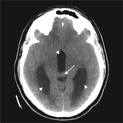
CT of the brain showing a small mass in the region of the pineal gland (arrow) with enlargement of the third and lateral ventricles (arrowhead).
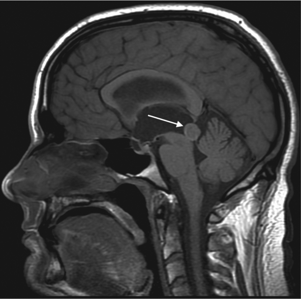
Precontrast sagittal T1-weighted MRI showing a mass in the region of the pineal gland, with a thin rim of intrinsic signal hyperintensity (arrow).
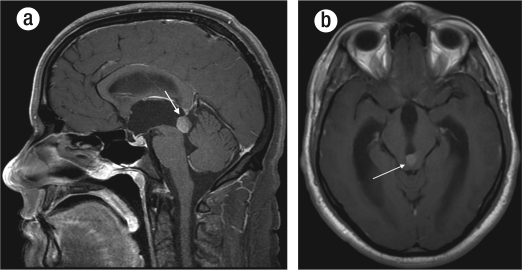
Postcontrast (a) sagittal and (b) axial T1-weighted images showing an avidly enhancing mass (arrows).
Similar articles
-
Papillary Tumor of the Pineal Region: MR Signal Intensity Correlated to Histopathology.Case Rep Neurol Med. 2015;2015:315095. doi: 10.1155/2015/315095. Epub 2015 Jan 22. Case Rep Neurol Med. 2015. PMID: 25688307 Free PMC article.
-
Nuclear CRX and FOXJ1 Expression Differentiates Non-Germ Cell Pineal Region Tumors and Supports the Ependymal Differentiation of Papillary Tumor of the Pineal Region.Am J Surg Pathol. 2017 Oct;41(10):1410-1421. doi: 10.1097/PAS.0000000000000903. Am J Surg Pathol. 2017. PMID: 28719464
-
Papillary tumors of the pineal region: case report.Neurosurgery. 2007 May;60(5):E953-5; discussion E953-5. doi: 10.1227/01.NEU.0000255443.44365.77. Neurosurgery. 2007. PMID: 17460510
-
Papillary tumor of the pineal region: Histopathological characterization and review of the literature.Neurochirurgie. 2015 Apr-Jun;61(2-3):138-42. doi: 10.1016/j.neuchi.2013.04.011. Epub 2014 Feb 18. Neurochirurgie. 2015. PMID: 24556386 Review.
-
Histopathology of tumors of the pineal region.Future Oncol. 2010 May;6(5):791-809. doi: 10.2217/fon.10.28. Future Oncol. 2010. PMID: 20465391 Review.
Cited by
-
Role of surgery, radiotherapy and chemotherapy in papillary tumors of the pineal region: a multicenter study.J Neurooncol. 2013 Apr;112(2):223-31. doi: 10.1007/s11060-013-1050-5. Epub 2013 Jan 12. J Neurooncol. 2013. PMID: 23314823
-
Pineal Gland Tumors: A Review.Cancers (Basel). 2021 Mar 27;13(7):1547. doi: 10.3390/cancers13071547. Cancers (Basel). 2021. PMID: 33801639 Free PMC article. Review.
-
Papillary tumor of the pineal region: A case report and review of the literature.Exp Ther Med. 2015 Oct;10(4):1375-1379. doi: 10.3892/etm.2015.2696. Epub 2015 Aug 20. Exp Ther Med. 2015. PMID: 26622493 Free PMC article.
-
Determination of Optimal Treatment Plan for Papillary Tumor of the Pineal Region: Case Series With Literature Review.Brain Tumor Res Treat. 2024 Oct;12(4):221-229. doi: 10.14791/btrt.2024.0021. Brain Tumor Res Treat. 2024. PMID: 39542518 Free PMC article.
References
-
- Smith AB, Rushing EJ, Smirniotopoulos JG. From the archives of the AFIP: Lesions of the pineal region: radiologic-pathologic correlation. Radiographics. 2010;30(7):2001–2020. - PubMed
-
- Kim YH, Kim JW, Park CK, Kim DG, Sohn CH, Chang KH, Park SH. Papillary tumor of pineal region presenting with leptomeningeal seeding. Neuropathology. 2010;30(6):654–660. - PubMed
-
- Kaloshi G, Rroji A, Lame A, Leka L, Haxhihyseni E, Vreto G, Petrela M. Natural history of papillary tumor of the pineal region: new insights on biological explanation. J Neurooncol. 2010;100(3):487–488. - PubMed
LinkOut - more resources
Full Text Sources
Research Materials