The P2X(7) receptor regulates proteoglycan expression in the corneal stroma
- PMID: 22275804
- PMCID: PMC3265178
The P2X(7) receptor regulates proteoglycan expression in the corneal stroma
Abstract
Purpose: Previously, the authors demonstrated that the lack of the P2X(7) receptor impairs epithelial wound healing and stromal collagen organization in the cornea. The goal here is to characterize specific effects of the P2X(7) receptor on components of the corneal stroma extracellular matrix.
Methods: Unwounded corneas from P2X(7) knockout mice (P2X(7) (-/-)) and C57BL/6J wild type mice (WT) were fixed and prepared for quantitative and qualitative analysis of protein expression and localization using Real Time PCR and immunohistochemistry. Corneas were stained also with Cuprolinic blue for electron microscopy to quantify proteoglycan sulfation in the stroma.
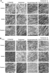
Results: P2X(7) (-/-) mice showed decreased mRNA expression in the major components of the corneal stroma: collagen types I and V and small leucine-rich proteoglycans decorin, keratocan, and lumican. In contrast P2X(7) (-/-) mice showed increased mRNA expression in lysyl oxidase and biglycan. Additionally, we observed increases in syndecan 1, perlecan, and type III collagen. There was a loss of perlecan along the basement membrane and enhanced expression throughout the stroma, in contrast with the decreased localization of other proteoglycans throughout the stroma. In the absence of lyase digestion there was a significantly smaller number of proteoglycan units per 100 nm of collagen fibrils in the P2X(7) (-/-) compared to WT mice. While digestion was more pronounced in the WT group, double digestion with Keratanase I and Chondroitinase ABC removed 88% of the GAG filaments in the WT, compared to 72% of those in the P2X(7) (-/-) mice, indicating that there are more heparan sulfate proteoglycans in the latter.
Conclusions: Our results indicate that loss of P2X(7) alters both the expression of proteins and the sulfation of proteoglycans in the corneal stroma.
Figures


Similar articles
-
Regulation by P2X7: epithelial migration and stromal organization in the cornea.Invest Ophthalmol Vis Sci. 2008 Oct;49(10):4384-91. doi: 10.1167/iovs.08-1688. Epub 2008 May 23. Invest Ophthalmol Vis Sci. 2008. PMID: 18502993 Free PMC article.
-
Interclass small leucine-rich repeat proteoglycan interactions regulate collagen fibrillogenesis and corneal stromal assembly.Matrix Biol. 2014 Apr;35:103-11. doi: 10.1016/j.matbio.2014.01.004. Epub 2014 Jan 18. Matrix Biol. 2014. PMID: 24447998 Free PMC article.
-
Altered KSPG expression by keratocytes following corneal injury.Mol Vis. 2003 Nov 21;9:615-23. Mol Vis. 2003. PMID: 14654769
-
The molecular basis of corneal transparency.Exp Eye Res. 2010 Sep;91(3):326-35. doi: 10.1016/j.exer.2010.06.021. Epub 2010 Jul 3. Exp Eye Res. 2010. PMID: 20599432 Free PMC article. Review.
-
Roles of lumican and keratocan on corneal transparency.Glycoconj J. 2002 May-Jun;19(4-5):275-85. doi: 10.1023/A:1025396316169. Glycoconj J. 2002. PMID: 12975606 Review.
Cited by
-
Hypoxia modulates the development of a corneal stromal matrix model.Exp Eye Res. 2018 May;170:127-137. doi: 10.1016/j.exer.2018.02.021. Epub 2018 Feb 27. Exp Eye Res. 2018. PMID: 29496505 Free PMC article.
-
Animal Models for the Investigation of P2X7 Receptors.Int J Mol Sci. 2023 May 4;24(9):8225. doi: 10.3390/ijms24098225. Int J Mol Sci. 2023. PMID: 37175933 Free PMC article. Review.
-
The Role of the Lysyl Oxidases in Tissue Repair and Remodeling: A Concise Review.Tissue Eng Regen Med. 2017 Jan 17;14(1):15-30. doi: 10.1007/s13770-016-0007-0. eCollection 2017 Feb. Tissue Eng Regen Med. 2017. PMID: 30603458 Free PMC article. Review.
-
Glycosaminoglycans: Roles in wound healing, formation of corneal constructs and synthetic corneas.Ocul Surf. 2023 Oct;30:85-91. doi: 10.1016/j.jtos.2023.08.008. Epub 2023 Aug 30. Ocul Surf. 2023. PMID: 37657650 Free PMC article. Review.
-
Hyperosmotic stress induces ATP release and changes in P2X7 receptor levels in human corneal and conjunctival epithelial cells.Purinergic Signal. 2017 Jun;13(2):249-258. doi: 10.1007/s11302-017-9556-5. Epub 2017 Feb 7. Purinergic Signal. 2017. PMID: 28176024 Free PMC article.
References
-
- Baraldi PG, Di Virgilio F, Romagnoli R. Agonists and antagonists acting at P2X7 receptor. Curr Top Med Chem. 2004;4:1707–17. - PubMed
-
- Wang Q, Wang L, Feng Y-H, Li X, Zeng R, Gorodeski GI. P2X7 receptor-mediated apoptosis of human cervical epithelial cells. Am J Physiol Cell Physiol. 2004;287:C1349–58. - PubMed
-
- Budagian V, Bulanova E, Brovko L, Orinska Z, Fayad R, Paus R, Bulfone-Paus S. Signaling through P2X7 Receptor in Human T Cells Involves p56 lck, MAP Kinases, and Transcription Factors AP-1 and NF-κB. J Biol Chem. 2003;278:1549–60. - PubMed
Publication types
MeSH terms
Substances
Grants and funding
LinkOut - more resources
Full Text Sources
Medical
Molecular Biology Databases
