

Neuroimaging in anxiety disorders
- PMID: 22275850
- PMCID: PMC3263392
- DOI: 10.31887/DCNS.2011.13.4/kholzschneider
Neuroimaging in anxiety disorders
Abstract
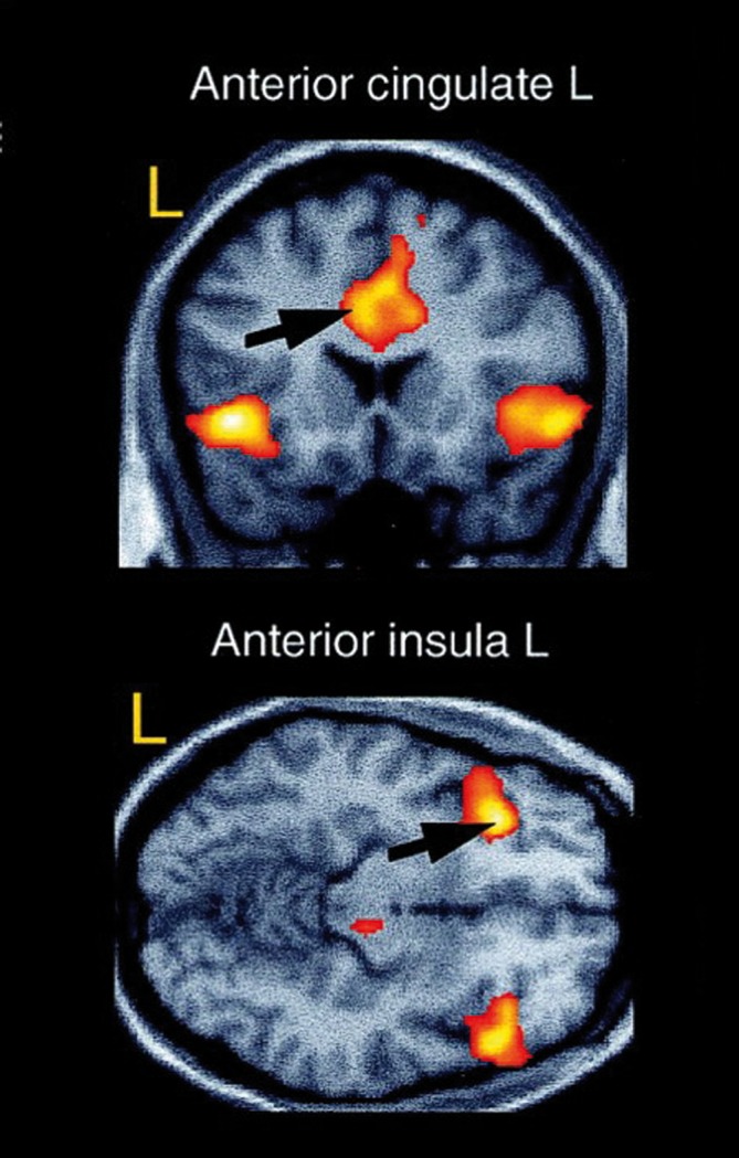
Over the last few years, neuroimaging techniques have contributed greatly to the identification of the structural and functional neuroanatomy of anxiety disorders. The amygdala seems to be a crucial structure for fear and anxiety, and has consistently been found to be activated in anxiety-provoking situations. Apart from the amygdala, the insula and anterior cinguiate cortex seem to be critical, and ail three have been referred to as the "fear network." In the present article, we review the main findings from three major lines of research. First, we examine human models of anxiety disorders, including fear conditioning studies and investigations of experimentally induced panic attacks. Then we turn to research in patients with anxiety disorders and take a dose look at post-traumatic stress disorder and obsessive-compulsive disorder. Finally, we review neuroimaging studies investigating neural correlates of successful treatment of anxiety, focusing on exposure-based therapy and several pharmacological treatment options, as well as combinations of both.
Durante los últimos años las técnicas de neuroimágenes han contribuido de manera importante a la identificación de la neuroanatomía estructural y funcional de los trastornos ansiosos. La amígdala parece ser una estructura crucial para el miedo y la ansiedad, y constantemente se ha encontrado activada en situaciones que provocan ansiedad, Además de la amígdala, la ínsula y la corieza cingulada anterior también parecen ser muy importantes y las tres se han denominado “el circuito del miedo”. En este artículo se revisan los principales hallazgos de tres importantes líneas de investigation. Primero se examinan modelos humanos de los trastornos ansiosos, incluyendo estudios de miedo condicionado e investigaciones de ataques de pánico inducidos experimenialmente. Luego se aborda la investigación en patientes con trastornos ansiosos con especial énfasis en el trastorno por estrés postraumático y el trastorno obsesivo compulsivo. Finalmente se revisan los estudios de neuroimágenes que investigan los correlatos neurales de tratamientos ansiolíticos exitosos, enfocándose en terapias basadas en la exposición y en algunas alternativas psicofarmacológicas, como también en combinaciones de ambas.
Ces dernières années, les techniques de neuro-imagerie ont largement contribué à l'identification de la neuroanatomie structurale et fonctionnelle des troubles anxieux. Les amygdales, structures capitales pour la peur et l'anxiété, sont régulièrement activées dans des situations pourvoyeuses d'anxiété, À côté des amygdales, l'insula et le cortex cingulaire antérieur semblent d'une importance cruciale, ces trois structures ayant été qualifiées de « réseau de la peur ». Dans cet article, nous passons en revue les principaux résultats de trois axes majeurs de recherche. Tout d'abord, nous examinons des modèles humains de troubles anxieux, à l'aide d'études sur le conditionnement de la peur et d'enquêtes sur les attaques de panique induites expérimentalement. Puis nous nous consacrons à la recherche chez les patients atteints de troubles anxieux et nous examinons de près l'état de stress post-traumatique et les troubles obsessionnels compulsifs. Enfin, nous analysons des études de neuroimagerie sur les corrélations neurales du traitement efficace de l'anxiété, en nous concentrant sur un traitement basé sur l'exposition (au stimulus anxiogène) et sur plusieurs traitements pharmacologiques, comme sur l'association des deux.
Keywords: amygdala; anxiety disorder; functional magnetic resonance imaging; neuroimaging; treatment.
Figures
Similar articles
-
Functional neuroimaging of anxiety: a meta-analysis of emotional processing in PTSD, social anxiety disorder, and specific phobia.Am J Psychiatry. 2007 Oct;164(10):1476-88. doi: 10.1176/appi.ajp.2007.07030504. Am J Psychiatry. 2007. PMID: 17898336 Free PMC article.
-
Neuroimaging studies of amygdala function in anxiety disorders.Ann N Y Acad Sci. 2003 Apr;985:389-410. doi: 10.1111/j.1749-6632.2003.tb07096.x. Ann N Y Acad Sci. 2003. PMID: 12724173 Review.
-
Functional imaging of mood and anxiety disorders.J Neuroimaging. 2006 Jan;16(1):1-10. doi: 10.1177/1051228405001474. J Neuroimaging. 2006. PMID: 16483270 Review.
-
Functional neuroimaging studies in posttraumatic stress disorder: review of current methods and findings.Depress Anxiety. 2007;24(3):202-18. doi: 10.1002/da.20208. Depress Anxiety. 2007. PMID: 16960853 Free PMC article. Review.
-
The human dimension: how the prefrontal cortex modulates the subcortical fear response.Rev Neurosci. 2007;18(3-4):191-207. doi: 10.1515/revneuro.2007.18.3-4.191. Rev Neurosci. 2007. PMID: 18019606 Review.
Cited by
-
Heritability estimation of subcortical volumes in a multi-ethnic multi-site cohort study.bioRxiv [Preprint]. 2024 Jan 12:2024.01.11.575231. doi: 10.1101/2024.01.11.575231. bioRxiv. 2024. PMID: 38260520 Free PMC article. Preprint.
-
Sliding Scale Theory of Attention and Consciousness/Unconsciousness.Behav Sci (Basel). 2022 Feb 10;12(2):43. doi: 10.3390/bs12020043. Behav Sci (Basel). 2022. PMID: 35200294 Free PMC article.
-
Neural correlates of a computerized attention modification program in anxious subjects.Soc Cogn Affect Neurosci. 2014 Sep;9(9):1379-87. doi: 10.1093/scan/nst128. Epub 2013 Aug 9. Soc Cogn Affect Neurosci. 2014. PMID: 23934417 Free PMC article.
-
Functional MRI in the investigation of blast-related traumatic brain injury.Front Neurol. 2013 Mar 4;4:16. doi: 10.3389/fneur.2013.00016. eCollection 2013. Front Neurol. 2013. PMID: 23460082 Free PMC article.
-
The Relationship Between the Uncinate Fasciculus and Anxious Temperament Is Evolutionarily Conserved and Sexually Dimorphic.Biol Psychiatry. 2019 Dec 15;86(12):890-898. doi: 10.1016/j.biopsych.2019.07.022. Epub 2019 Jul 31. Biol Psychiatry. 2019. PMID: 31542153 Free PMC article.
References
-
- Damsa C., Kosel M., Moussally J. Current status of brain imaging in anxiety disorders. Curr Opin Psychiatry. 2009;22:96–110. - PubMed
-
- American Psychiatric Association. Diagnostic and Statistical Manual of Mental Disorders. 4th ed. Text Revision. Washington, DC: American Psychiatric Association; 2000
-
- Buchel C., Morris J., Dolan RJ., Friston KJ. Brain systems mediating aversive conditioning: an event-related fMRI study. Neuron. 1998;20:947–957. - PubMed
Publication types
MeSH terms
LinkOut - more resources
Full Text Sources
Medical