. 2012 Jan;8(1):e1002483.
doi: 10.1371/journal.ppat.1002483. Epub 2012 Jan 19.

Impaired chromatin remodelling at STAT1-regulated promoters leads to global unresponsiveness of Toxoplasma gondii-infected macrophages to IFN-γ

Affiliations

Impaired chromatin remodelling at STAT1-regulated promoters leads to global unresponsiveness of Toxoplasma gondii-infected macrophages to IFN-γ

Christine Lang et al. PLoS Pathog. 2012 Jan.

Abstract

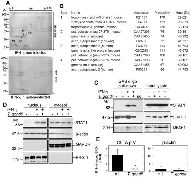
Intracellular pathogens including the apicomplexan and opportunistic parasite Toxoplasma gondii profoundly modify their host cells in order to establish infection. We have shown previously that intracellular T. gondii inhibit up-regulation of regulatory and effector functions in murine macrophages (MΦ) stimulated with interferon (IFN)-γ, which is the cytokine crucial for controlling the parasites' replication. Using genome-wide transcriptome analysis we show herein that infection with T. gondii leads to global unresponsiveness of murine macrophages to IFN-γ. More than 61% and 89% of the transcripts, which were induced or repressed by IFN-γ in non-infected MΦ, respectively, were not altered after stimulation of T. gondii-infected cells with IFN-γ. These genes are involved in a variety of biological processes, which are mostly but not exclusively related to immune responses. Analyses of the underlying mechanisms revealed that IFN-γ-triggered nuclear translocation of STAT1 still occurred in Toxoplasma-infected MΦ. However, STAT1 bound aberrantly to oligonucleotides containing the IFN-γ-responsive gamma-activated site (GAS) consensus sequence. Conversely, IFN-γ did not induce formation of active GAS-STAT1 complexes in nuclear extracts from infected MΦ. Mass spectrometry of protein complexes bound to GAS oligonucleotides showed that T. gondii-infected MΦ are unable to recruit non-muscle actin to IFN-γ-responsive DNA sequences, which appeared to be independent of stimulation with IFN-γ and of STAT1 binding. IFN-γ-induced recruitment of BRG-1 and acetylation of core histones at the IFN-γ-regulated CIITA promoter IV, but not β-actin was diminished by >90% in Toxoplasma-infected MΦ as compared to non-infected control cells. Remarkably, treatment with histone deacetylase inhibitors restored the ability of infected macrophages to express the IFN-γ regulated genes H2-A/E and CIITA. Taken together, these results indicate that Toxoplasma-infected MΦ are unable to respond to IFN-γ due to disturbed chromatin remodelling, but can be rescued using histone deacetylase inhibitors.


Conflict of interest statement

The authors have declared that no competing interests exist.

Figures

Figure 1
Figure 1. Regulation of the transcriptome of primary murine macrophages after infection with T. gondii and/or activation with IFN-γ.
(A) Primary BMMΦ were infected or not with T. gondii and 2 hours later, were stimulated with IFN-γ for additional 22 hours or left untreated. RNA from four biological replicates was used for further analysis. (B) After reverse transcription of mRNA and labelling with Cy3 or Cy5, two samples each were hybridized to mouse whole genome microarrays in a dye-swap loop design as indicated by arrows. (C) Number of probes and corresponding number of genes or transcripts that were at least 4-fold regulated at ≤ 1% FDR.
Figure 2
Figure 2. T. gondii infection severely impairs the ability of murine macrophages to respond to IFN-γ.
(A) The cDNAs from non-infected and T. gondii-infected murine BMMΦ stimulated or not with IFN-γ were hybridized to mouse whole genome microarrays. Microarray probes were identified, which showed at least 4-fold up- or down-regulation in IFN-γ-treated, non-infected cells versus untreated, non-infected cells. Expression levels of the corresponding genes in non-infected (n.i.) and T. gondii-infected cells before and after IFN-γ treatment are displayed following Z-score transformation using a blue (induced) to yellow (repressed) scale. (B) Out of the 41,174 good quality spots, those which were differentially regulated in at least one of the five comparisons as depicted in Figure 1B were selected (threshold 4-fold regulation; identified by ANOVA; n = 3527). They were subjected to a correlation analysis of the fold change of mRNA from non-infected control cells in response to IFN-γ, and the impact exerted by T. gondii on IFN-γ-regulated gene expression. Vertical lines at 0.25 and 4 indicate lower and upper thresholds for differentially expressed genes. The correlation coefficient r of the linear regression is indicated.
Figure 3
Figure 3. Impact of Toxoplasma infection on the IFN-γ-regulated transcriptome of murine BMMΦ as determined by mouse cDNA microarray analyses.
(A) Oligonucleotide probes on the array that were at least 4-fold up-regulated (IFN > Ctr) or 4-fold down-regulated (IFN < Ctr) after IFN-γ treatment of non-infected BMMΦ, as compared to untreated controls were identified at a FDR ≤ 1%, and were compared with un-regulated probes (IFN  =  Ctr). Total numbers of probes are given at the top of each bar. The percentages of probes of which the up- or down-regulation in non-infected cells was abrogated in Toxoplasma-infected cells (infected, IFN-γ-treated vs. infected, untreated) are indicated by black and cross-hatched bars, respectively. (B) Genes that were at least 4-fold up-regulated after IFN-γ-treatment of non-infected (black bars) or Toxoplasma-infected BMMΦ (cross-hatched bars) were clustered in biological processes using the DAVID Bioinformatics resource. Depicted are major IFN-γ-regulated clusters containing a minimum of 10 different genes, the expression of which was at least 4-fold induced after treating non-infected macrophages with IFN-γ. (C) Genes that were at least 4-fold down-regulated by IFN-γ-treatment of non-infected (black bars) or Toxoplasma-infected BMMΦ (cross-hatched bars) were clustered in biological processes using the DAVID Bioinformatics resource. Depicted are major IFN-γ-regulated clusters containing a minimum of 10 different genes, the expression of which was at least 4-fold repressed after treating non-infected macrophages with IFN-γ.
Figure 4
Figure 4. Binding activity of STAT1 homodimers to GAS-containing DNA sequences is modulated after Toxoplasma infection.
Murine BMMΦ were infected or not with T. gondii for 24 hours, and were stimulated with IFN-γ during the final 30 minutes, or were left non-stimulated. Nuclear extracts were tested for their binding to radiolabelled GAS-containing oligonucleotides by electromobility shift assay. Supershift assays were performed using antibodies against STAT1α, STAT2 or p48, or with normal rabbit IgG as a control. GAF: gamma activated factor.
Figure 5
Figure 5. Toxoplasma infection leads to defective binding of components of chromatin-remodelling complexes to GAS-containing oligonucleotides in vitro and to the IFN-γ responsive CIITA promoter IV in infected cells.
(A) RAW264.7 macrophages were infected with T. gondii for 24 hours, or were left non-infected and stimulated with IFN-γ during the final 30 min. Complete cellular lysates were used to isolate GAS-binding protein complexes by a pull-down assay. Precipitates were resolved by isoelectric focussing and SDS-PAGE, and proteins were visualized by silver staining. Protein spots that were specifically identified in the pull-downs from IFN-γ-treated, non-infected MΦ are indicated. (B) GAS-binding proteins that were pulled down from lysates of non-infected, but not Toxoplasma-infected MΦ after stimulation with IFN-γ were analysed by mass spectrometry. The three hits of highest probability for each of the four protein spots are shown; probability scores greater than 61 are considered significant. (C) RAW264.7 macrophages infected or not with T. gondii for 24 hours were stimulated with IFN-γ during the final 30 min, or were left non-stimulated. Complete cellular extracts or proteins that had been pulled-down from the extracts, by using a biotin-conjugated GAS-containing oligonucleotide and immobilized streptavidin, were subsequently analysed by immunoblotting using antibodies directed against STAT1, β-actin and BRG-1. Nc: negative control: pull-down assay without lysate. (D) Nuclear and cytosolic extracts were prepared from RAW264.7 macrophages infected with T. gondii, and stimulated with IFN-γ as described in (C). After separation of the extracts by SDS-PAGE, STAT1, β-actin, GAPDH and BRG-1 were detected by immunoblotting. (E) Toxoplasma-infected and non-infected (n.i.) RAW264.7 macrophages were stimulated with IFN-γ for 2 hours, or were left non-stimulated. After cross-linking DNA-protein complexes and shearing, chromatin was immunoprecipitated using an anti-BRG-1 antibody and protein A agarose. After DNA isolation from immunoprecipitates or from input lysates, fragments of the CIITA promoter IV (pIV) or β-actin were amplified by real-time PCR. The IFN-γ-induced binding of BRG-1 was calculated according to the ratio (EChIP)ΔCPChIP(unstimulated – IFN-γ-treated)/(Einput)ΔCPinput(unstimulated – IFN-γ-treated). Results are means ± S.E.M. (n = 2), significant differences were identified by Student's t-test (*, p<0.05).
Figure 6
Figure 6. Toxoplasma infection impairs IFN-γ-induced acetylation of histones H3 and H4 at IFN-γ-responsive promoters in murine macrophages.
(A) RAW264.7 macrophages were infected with T. gondii for 24 hours or were left non-infected, and were stimulated or not with IFN-γ during the final 16 hours. After cross-linking DNA-protein complexes, cell lysates were subjected to ChIP analysis using an anti-acetyl-H4 antibody and protein A agarose. A no-antibody control was run in parallel (wo Ab). After isolation of DNA from chromatin immunoprecipitates or from input cell lysates, fragments of the CIITA pIV or β-actin were amplified by real-time PCR. Amplicons were verified by agarose gel electrophoresis. Nc: PCR negative control: without template. (B+C) Acetylation of histones H4 (B) and H3 (C) at the CIITA pIV and β-actin after stimulation of MΦ with IFN-γ was analysed by ChIP as described above and was quantified according to the ratio (EChIP)ΔCPChIP(unstimulated – IFN-γ-treated)/(Einput)ΔCPinput(unstimulated – IFN-γ-treated). Results are means ± S.E.M. from three independent experiments, significant differences between the acetylation in non-infected (n.i.) and infected (T. gondii) MΦ were identified by Student's t-test (**, p<0.01). (D) The histone H3/H4 acetylation at the H2-Eβ and the GBP2 promoters of non-infected and Toxoplasma-infected MΦ was analysed by ChIP as described above (n = 2).
Figure 7
Figure 7. HDAC inhibitors restore responsiveness of Toxoplasma-infected macrophages to IFN-γ.
(A) RAW264.7 macrophages were infected with T. gondii (parasite-host cell ratio 3∶1), or were left non-infected and stimulated with IFN-γ in the absence (upper panel) or presence of 2 µM of the HDAC inhibitor MS-275 (lower panel). Forty hours after infection, cells were immunolabelled with anti-H2-A/E and analysed by flow cytometry. Results are from a representative experiment out of three, data are the percentages of cells within the individual quadrants. (B) Murine macrophages were infected with T. gondii and/or treated with IFN-γ and MS-275 as described in (A). Cells were immunolabelled with an antibody directed against H2-A/E or an isotype-matched control antibody, and were analysed by flow cytometry (please consider the different scaling of the x-axes in both plots). Data represent the mean percentages ± S.E.M. of positive cells from three independent experiments. *: p<0.05, **: p<0.01, ns: not significant (Student's t-test). (C) IFN-γ-induced transcript levels of CIITA and H2-Aβ in infected and non-infected RAW264.7 macrophages after treatment with HDAC inhibitors MS-275 or sodium butyrate. Cells were infected or not with T. gondii for 24 hours, and treated with IFN-γ in the absence (control) or in the presence of 0.5 or 2 µM sodium butyrate or 0.5, 1 or 2 µM MS-275 as indicated. After isolation of RNA and reverse transcription of mRNA, CIITA, H2-Aβ and β-actin were amplified, and semi-quantitatively analysed. IFN-γ-induced CIITA and H2-Aβ transcript levels in T. gondii-infected and non-infected samples were normalized to β-actin, and the fold inhibition imposed by parasitic infection was calculated. Data represent means ± S.E.M. from three independent experiments. (D) Impact of HDAC inhibitors targeting different HDACs on Toxoplasma-mediated inhibition of H2-A/E expression. Murine macrophages were infected with T. gondii and/or treated with IFN-γ and MS-275, MC1568 (both at 2 µM) or nicotinamide (5 mM) as described in (A). After immunolabelling with an antibody directed against H2-A/E, cells were analysed by FACS. The parasite-imposed inhibition of IFN-γ-induced H2-A/E expression was calculated; data represent means ± S.E.M. from three independent experiments. (E) Acetylation of histone H4 at the CIITA pIV in the presence or absence of MS-275. Toxoplasma-infected and non-infected MΦ (n.i.) were treated or not with 2 µM MS-275, and stimulated with IFN-γ or left non-stimulated. After cross-linking DNA-protein complexes, cell lysates were analysed by ChIP using an anti-acetyl-H4 antibody. ChIP and input DNA were amplified by real-time PCR using primers specific for the CIITA pIV. The IFN-γ-induced H4 acetylation in the presence or absence of MS-275 was quantified according to the ratio (EChIP)ΔCPChIP(unstimulated – IFN-γ-treated)/(Einput)ΔCPinput(unstimulated – IFN-γ-treated). Results are means ± S.E.M. from two independent experiments.
Figure 8
Figure 8. Model of Toxoplasma-imposed inhibition of IFN-γ-mediated gene expression, and the rescue of murine MΦ from such unresponsiveness using HDAC inhibitors.
In the absence of IFN-γ, hypoacetylated histones due to predominant HDAC activity lead to compact chromatin and repression of promoter activation (upper left). After stimulation with IFN-γ, recruitment of BAF including the ATPase BRG-1 leads to changes in chromatin structure that allows subsequent binding of STAT1 to its consensus sequence, recruitment of HATs, and hyperacetylation of histones and non-histone proteins. Recruitment of BAF as well as full activity of BRG-1 and possibly other components of active STAT1-regulated promoters require the presence of non-muscle actin. Finally, binding of IRF-1 allows binding of the transcriptional machinery and Pol II-driven gene transcription (upper right). In Toxoplasma-infected MΦ, reduced levels of actin at STAT1-responsive promoters coincide with reduced recruitment of chromatin remodelling complexes, including BRG-1 and HATs and thereby favour hypoacetylated chromatin. STAT1 does either aberrantly bind to such promoters, or does not bind in infected cells (lower left). Treatment of Toxoplasma-infected MΦ with HDAC inhibitors leads to a shift towards increased acetylation of histones and/or non-histone proteins, and thereby favours the switch to permissive chromatin following IFN-γ activation. This facilitates binding of transcription factors and chromatin remodellers in the presence of T. gondii, and allows binding of the transcriptional machinery and IFN-γ-induced gene expression (lower right).

Similar articles

Cited by

References

    1. Montoya JG, Liesenfeld O. Toxoplasmosis. Lancet. 2004;363:1965–1976. - PubMed
    1. Lüder CG, Stanway RR, Chaussepied M, Langsley G, Heussler VT. Intracellular survival of apicomplexan parasites and host cell modification. Int J Parasitol. 2009;39:163–173. - PubMed
    1. Suzuki Y, Orellana MA, Schreiber RD, Remington JS. Interferon-gamma: the major mediator of resistance against Toxoplasma gondii. Science. 1988;240:516–518. - PubMed
    1. Nathan CF, Murray HW, Wiebe ME, Rubin BY. Identification of interferon-gamma as the lymphokine that activates human macrophage oxidative metabolism and antimicrobial activity. J Exp Med. 1983;158:670–689. - PMC - PubMed
    1. Gazzinelli RT, Hakim FT, Hieny S, Shearer GM, Sher A. Synergistic role of CD4+ and CD8+ T lymphocytes in IFN-gamma production and protective immunity induced by an attenuated Toxoplasma gondii vaccine. J Immunol. 1991;146:286–292. - PubMed

Publication types

MeSH terms