. 2011 Nov 24:5:94.
doi: 10.3389/fnsys.2011.00094. eCollection 2011.

Extracellular glutamate: functional compartments operate in different concentration ranges

Affiliations

Extracellular glutamate: functional compartments operate in different concentration ranges

Khaled Moussawi et al. Front Syst Neurosci. .

Abstract

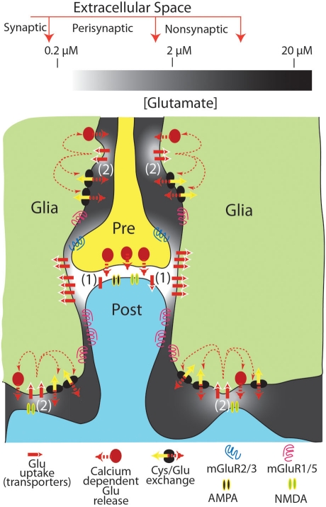
Extracellular glutamate of glial origin modulates glial and neuronal glutamate release and synaptic plasticity. Estimates of the tonic basal concentration of extracellular glutamate range over three orders of magnitude (0.02-20 μM) depending on the technology employed to make the measurement. Based upon binding constants for glutamate receptors and transporters, this range of concentrations translates into distinct physiological and pathophysiological roles for extracellular glutamate. Here we speculate that the difference in glutamate measurements can be explained if there is patterned membrane surface expression of glutamate release and transporter sites creating extracellular subcompartments that vary in glutamate concentration and are preferentially sampled by different technologies.

Keywords: cystine–glutamate exchange; glia; glutamate; glutamate uptake; mGluR; synapse.


Figures

Figure 1
Figure 1
Tonic levels of extracellular glutamate measured in the synaptic, perisynaptic, and nonsynaptic compartments of the extracellular space. The levels of glutamate shown are approximated for resting conditions (i.e., in the absence of action potential mediated synaptic release). It is proposed that extracellular glutamate in compartmentalized morphologically by close appositions between neuronal and glial processes (note that the distances between glia–neuron distances are exaggerated for illustrative purposes), and by the patterned distribution of glutamate uptake sites and neuronal or glial release sites. This creates 2 regions of nanomolar glutamate high in iGluR, uptake sites and either synaptic (1) or glial (2) glutamate release sites. A second region surrounding the nanomolar region contains low micromolar levels of glutamate that are maintained largely from glial release and relatively lower glutamate uptake via high affinity transporters. This latter compartment is in part populated by mGluR that receive tonic glutamate stimulation and thereby modulate synaptic (and probably glial; D’Ascenzo et al., 2007) release and neuroplasticity.

References

    1. Acuna-Goycolea C., Li Y., van den Pol A. N. (2004). Group III metabotropic glutamate receptors maintain tonic inhibition of excitatory synaptic input to hypocretin/orexin neurons. J. Neurosci. 24, 3013–3022 10.1523/JNEUROSCI.5416-03.2004 - DOI - PMC - PubMed
    1. Aoyama K., Watabe M., Nakaki T. (2008). Regulation of neuronal glutathione synthesis. J. Pharmacol. Sci. 108, 227–238 10.1254/jphs.08R01CR - DOI - PubMed
    1. Baker D. A., McFarland K., Lake R. W., Shen H., Tang X. C., Toda S., Kalivas P. W. (2003). Neuroadaptations in cystine-glutamate exchange underlie cocaine relapse. Nat. Neurosci. 6, 743–749 10.1038/nn1069 - DOI - PubMed
    1. Baker D. A., Xi Z. X., Shen H., Swanson C. J., Kalivas P. W. (2002). The origin and neuronal function of in vivo nonsynaptic glutamate. J. Neurosci. 22, 9134–9141 - PMC - PubMed
    1. Bergles D. E., Diamond J. S., Jahr C. E. (1999). Clearance of glutamate inside the synapse and beyond. Curr. Opin. Neurobiol. 9, 293–298 10.1016/S0959-4388(99)80043-9 - DOI - PubMed

LinkOut - more resources