

An in vivo MRI Template Set for Morphometry, Tissue Segmentation, and fMRI Localization in Rats
- PMID: 22275894
- PMCID: PMC3254174
- DOI: 10.3389/fninf.2011.00026
An in vivo MRI Template Set for Morphometry, Tissue Segmentation, and fMRI Localization in Rats
Abstract
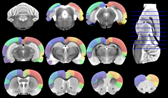
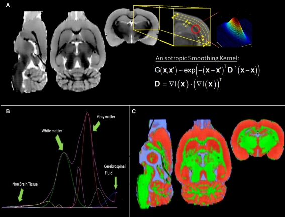
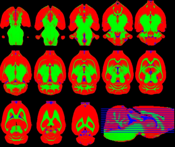
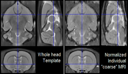
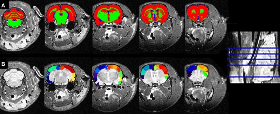
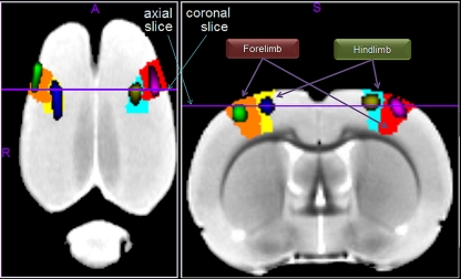
Over the last decade, several papers have focused on the construction of highly detailed mouse high field magnetic resonance image (MRI) templates via non-linear registration to unbiased reference spaces, allowing for a variety of neuroimaging applications such as robust morphometric analyses. However, work in rats has only provided medium field MRI averages based on linear registration to biased spaces with the sole purpose of approximate functional MRI (fMRI) localization. This precludes any morphometric analysis in spite of the need of exploring in detail the neuroanatomical substrates of diseases in a recent advent of rat models. In this paper we present a new in vivo rat T2 MRI template set, comprising average images of both intensity and shape, obtained via non-linear registration. Also, unlike previous rat template sets, we include white and gray matter probabilistic segmentations, expanding its use to those applications demanding prior-based tissue segmentation, e.g., statistical parametric mapping (SPM) voxel-based morphometry. We also provide a preliminary digitalization of latest Paxinos and Watson atlas for anatomical and functional interpretations within the cerebral cortex. We confirmed that, like with previous templates, forepaw and hindpaw fMRI activations can be correctly localized in the expected atlas structure. To exemplify the use of our new MRI template set, were reported the volumes of brain tissues and cortical structures and probed their relationships with ontogenetic development. Other in vivo applications in the near future can be tensor-, deformation-, or voxel-based morphometry, morphological connectivity, and diffusion tensor-based anatomical connectivity. Our template set, freely available through the SPM extension website, could be an important tool for future longitudinal and/or functional extensive preclinical studies.
Keywords: Paxinos and Watson; SPM; Wistar rats; anatomical connectivity; elastix; fMRI; morphometry; template set.
Figures
Similar articles
-
A reliable spatially normalized template of the human spinal cord--Applications to automated white matter/gray matter segmentation and tensor-based morphometry (TBM) mapping of gray matter alterations occurring with age.Neuroimage. 2015 Aug 15;117:20-8. doi: 10.1016/j.neuroimage.2015.05.034. Epub 2015 May 21. Neuroimage. 2015. PMID: 26003856
-
Construction and evaluation of multitracer small-animal PET probabilistic atlases for voxel-based functional mapping of the rat brain.J Nucl Med. 2006 Nov;47(11):1858-66. J Nucl Med. 2006. PMID: 17079820
-
Fully-integrated framework for the segmentation and registration of the spinal cord white and gray matter.Neuroimage. 2017 Apr 15;150:358-372. doi: 10.1016/j.neuroimage.2016.09.026. Epub 2016 Sep 20. Neuroimage. 2017. PMID: 27663988
-
[Understanding Voxel-Based Morphometry].Brain Nerve. 2017 May;69(5):505-511. doi: 10.11477/mf.1416200776. Brain Nerve. 2017. PMID: 28479527 Review. Japanese.
-
Structural brain atlases: design, rationale, and applications in normal and pathological cohorts.J Alzheimers Dis. 2012;31 Suppl 3(0 3):S169-88. doi: 10.3233/JAD-2012-120412. J Alzheimers Dis. 2012. PMID: 22647262 Free PMC article. Review.
Cited by
-
Topographical reorganization of brain functional connectivity during an early period of epileptogenesis.Epilepsia. 2021 May;62(5):1231-1243. doi: 10.1111/epi.16863. Epub 2021 Mar 15. Epilepsia. 2021. PMID: 33720411 Free PMC article.
-
Differences between normal and diabetic brains in middle-aged rats by MRI.Brain Res. 2019 Dec 1;1724:146407. doi: 10.1016/j.brainres.2019.146407. Epub 2019 Aug 26. Brain Res. 2019. PMID: 31465773 Free PMC article.
-
Development and advancements in rodent MRI-based brain atlases.Heliyon. 2024 Mar 8;10(6):e27421. doi: 10.1016/j.heliyon.2024.e27421. eCollection 2024 Mar 30. Heliyon. 2024. PMID: 38510053 Free PMC article. Review.
-
Data-Driven Regularization Parameter Selection in Dynamic MRI.J Imaging. 2021 Feb 20;7(2):38. doi: 10.3390/jimaging7020038. J Imaging. 2021. PMID: 34460637 Free PMC article.
-
Brain Morphometry and Longitudinal Relaxation Time of Spontaneously Hypertensive Rats (SHRs) in Early and Intermediate Stages of Hypertension Investigated by 3D VFA-SPGR MRI.Neuroscience. 2019 Apr 15;404:14-26. doi: 10.1016/j.neuroscience.2019.01.030. Epub 2019 Jan 26. Neuroscience. 2019. PMID: 30690138 Free PMC article.
References
-
- Alemán-Gómez Y., Melie-García L., Valdes-Hernández P. (2006). IBASPM: toolbox for automatic parcellation of brain structures. Presented at the 12th Annual Meeting of the Organization for Human Brain Mapping, June 11–15, 2006, Florence [Available on CD-Rom in NeuroImage, Vol. 27, No.1].
-
- Ashburner J. (2002). “Another MRI bias correction approach,” in Neuroimage 16, Presented at the 8th International Conference on Functional Mapping of the Human Brain, June 2–6, 2002, Sendai [Available on CD-Rom].
LinkOut - more resources
Full Text Sources