. 2012 Jan 11:2:281.
doi: 10.3389/fmicb.2011.00281. eCollection 2011.

Inhibition of the Plasma-Membrane-Associated Serine Protease Cathepsin G by Mycobacterium tuberculosis Rv3364c Suppresses Caspase-1 and Pyroptosis in Macrophages

Affiliations

Inhibition of the Plasma-Membrane-Associated Serine Protease Cathepsin G by Mycobacterium tuberculosis Rv3364c Suppresses Caspase-1 and Pyroptosis in Macrophages

Lia Danelishvili et al. Front Microbiol. .

Abstract

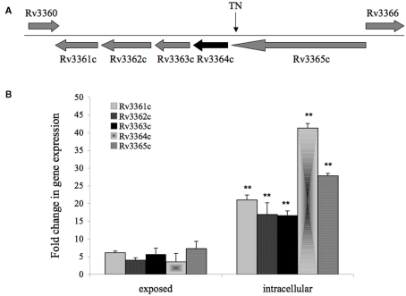
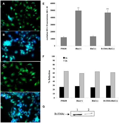
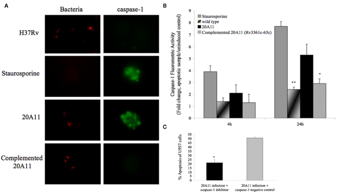
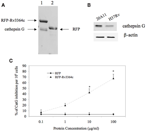
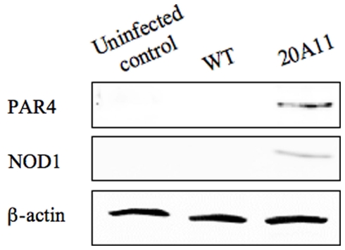
Tuberculosis is a disease associated with the infection of a great part of the world's population and is responsible for the death of two to three million people annually. Mycobacterium tuberculosis infects macrophages and subverts its mechanisms of killing. The pathogen suppresses macrophage apoptosis by many different mechanisms. We describe that, upon uptake by macrophages, M. tuberculosis overexpresses an operon Rv3361c-Rv3365c and secretes Rv3364c. The Rv3365c knockout strain is deficient in apoptosis inhibition. The Rv3364c protein binds to the serine protease cathepsin G on the membrane, inhibiting its enzymatic activity and the downstream activation of caspase-1-dependent apoptosis. In summary, M. tuberculosis prevents macrophage pyroptosis by a novel mechanism involving cytoplasmic surveillance proteins.

Keywords: Mycobacterium tuberculosis; apoptosis; caspase-1-dependent; inhibition; macrophages; pyroptosis.


Figures

Figure 1
Figure 1
(A) Gene organization of 20A11 mutant associated with induction of macrophage apoptosis. The operon consists of five genes, including histidine kinase sensor Rv3365c, possible structural or regulatory Rv3364c, DNA-binding Rv3363c, ATP/GTP binding Rv3362c, and putative Rv3361c that most likely forms pore. The site disrupted by the insertion of Tn5367 transposon (TN) is indicated with an arrow. (B) Real-Time PCR quantification of M. tuberculosis histidine kinase operon at a 30-min exposure or 4-h infection of macrophages. RNAs of intracellular or macrophage-exposed bacteria, and broth-grown bacteria were assessed to determine cDNA copy numbers for target and reference genes. Ct values were normalized with an internal housekeeping gene (16S) and fold change calculated between differentially expressed experimental and control transcripts, as previously reported (1). The data represent the average of three independent experiments ± SD. A p value of <0.01 indicated by ** was calculated between gene expressions in intracellular and exposed bacteria.
Figure 2
Figure 2
Mycobacterium tuberculosis Rv3364c protein translocation into cytoplasm of U937 cells visualized by β-lactamase-catalyzed hydrolysis of cell-permeable CCF2 (FRET-based) substrate. β-Lactamase hydrolysis in (A) macrophages infected with M. tuberculosis BlaC deletion clone PM638; (B) cells infected with M. tuberculosis clone expressing full length BlaC protein; (C) macrophages infected with PM638 clone containing BlaC(−) lacking a signal sequence; and (D) U937 cells infected with M. tuberculosis PM638 clone expressing Rv3364c:BlaC(−). While uncleaved CCF2 substrate emits green fluorescence, cleaved substrate by translocated β-lactamase from bacteria emits blue fluorescence. (E) The readings from CCF2-AM hydrolysis were also recorded using Infinity 200 cytofluorometer (Tecan). The values are means ± SD of three separate experiments performed in duplicate. **p < 0.01, The significance of differences between PM638 BlaC(+) and PM638; and between Rv3364c:BlaC(−) clone and PM638 BlaC(−). (F) Percentage of infected macrophages after 1 or 2 h of incubation as a function of the number of ingested PM638, PM638 BlaC(+), PM638 BlaC(−), or Rv3364c:BlaC(−) clone. Data from one experiment performed in duplicate and representative of three independent experiments. (G) Western blot analysis of Rv3364c protein in the cytoplasm of infected macrophages. U937 cells were infected with M tuberculosis containing pLDG13:His:Rv3364c over-expressed vector at an MOI 1:10. Over-expressed bacterial lysate and cell supernatants were subjected to immunoprecipitation and western blotting using His-tag antibody. Line 1, His:Rv3364c protein extracted from the over-expressed M. tuberculosis; Line 2, His:Rv3364c protein identified in the cytoplasmic fraction of macrophages.
Figure 3
Figure 3
(A)Production of IL-12, TNF, IFN-β, and TGF-β cytokines over time by macrophages infected with M. tuberculosis H37Rv and 20A11 mutant. Cells were infected with MOI of 10. The numbers represent the mean ± SD of three independent experiments. The significance of differences between the 20A11 mutant and the wild-type strain was evaluated *p < 0.05 and **p < 0.01 at the same time point infections. (B) Expression profile of intracellular pattern recognition receptor NOD1 and NOD2 and NF-κB in H37Rv- or 20A11-infected macrophages analyzed by Real-Time quantitative PCR. Bars represent the fold change between differentially expressed transcripts from wild-type and mutant infected cells. Positive values indicate relative increased expression levels over uninfected control. The numbers represent the mean ± SD of three independent experiments. The differences between the 20A11 mutant and H37Rv strain was evaluated *p < 0.05 and **p < 0.01 at the same times of post-infection. (C) Western blot analysis of NOD1 and NOD2 proteins. U937 cells were infected with M. tuberculosis H37Rv or 20A11 mutant (MOI 1:10) and cultured for 24 h and 4 days. Immunoprecipitated cell lysates were subjected to Western blotting, as described in Section “Materials and Methods.” (D) NOD1, NOD2 and β-actin protein levels were quantified via semi-quantitative Western blot analysis on the Li-Cor Odyssey Platform. The data represent the average of three independent experiments ± SD. A *p < 0.05 for NOD1 and NOD2 protein levels at day 4 compared with 24 h post-infection with 20A11 and H37Rv, respectively. **p < 0.01 Difference in NOD2 protein levels at day 4 compared with 24 h time post-infection with 20A11 mutant.
Figure 4
Figure 4
Role of caspase-1-dependent apoptosis. (A) In situ analysis of U937 macrophages for caspase-1 activation during M. tuberculosis H37Rv and 20A11 infection (MOI 10:1). While staurosporine treatment and 20A11 infection triggered caspase-1 activation of macrophages, M. tuberculosis H37Rv and complemented 20A11 clone were able to suppress caspase-1 activation. Bar, 10 μm. (B) M. tuberculosis infection prevents caspase-1 activation in macrophages. Cells were infected with wild-type or mutant strains, and fold change of caspase-1 activation was calculated according the manufacturer’s protocol. The data represent the average of three independent experiments ± SD. *p < 0.05 And **p < 0.01 statistically significant differences between complemented 20A11 and wild-type infection versus 20A11 mutant after 24 h post-infection, respectively. Active caspase-1 was quantified in situ using FAM–YVAD–FMK caspase-1 detection assay (Bachem). (C) Apoptosis level in caspase-1 inhibited macrophages after 24 h of 21A11 mutant post-infection examined by TUNEL. The values are means ± SD of two separate experiments performed in duplicate. The significance of differences between caspase-1 inhibitor and the caspase-1 negative control groups during 20A11 mutant infection was evaluated *p < 0.05.
Figure 5
Figure 5
Host target protein for Rv3364c. (A) The His-paramagnetic pre-charged nickel-pulling assay of macrophage proteins interacting with M. tuberculosis Rv3364c identified 29 kDa plasma-membrane-associated serine protease cathepsin G. (1) His-tag-RFP–Rv3364c pull-up sample with cathepsin G; (2) His-tag-RFP control pull-up sample. (B) Cathepsin G protein expression levels by Western blot analysis: (1) 20A11 mutant infection; (2) M. tuberculosis H37Rv infection. (C) The effects of the Rv3364c protein on cathepsin G activity. Purified His-tag-RFP–Rv3364c protein was exposed for 4 h to pre-cleared macrophage lysates at 0.1–100 μg/ml concentration range and changes in cathepsin G activity were measured as% of cathepsin G inhibition per 106 cells. The data represent the average of three independent experiments ± SD. *p < 0.05 for the comparison between His-tag-RFP–Rv3364c experimental and His-tag-RFP control treatments at the same concentration of proteins.
Figure 6
Figure 6
Colocalization of Rv3364c with the cathepsin G protein. U937 cells were transiently coinfected with Lenti-X viruses produced from a pLVX–ZsGreen1–cathepsin G vector and either a pLVX–dtTomato (A–D) or pLVX–dtTomato-Rv3364c fusion vector (E–H). ZsGgreen1-labeled cathepsin G visualization show punctate pattern in transduced cells (B,F). dtTomato labeled Rv3364c protein is also visible as punctate pattern (E), whereas dtTomato plane protein is diffused in the cytoplasm of macrophages (A). Nuclei are stained with DAPI (C,G). The overlap between cathepsin G and Rv3364c immunoreactivity appears yellow in the merged micrographs indicating colocalization of studied proteins (H). Scale bar, 10 μm.
Figure 7
Figure 7
Western blot analysis of PAR4 and NOD1 proteins. M. tuberculosis H37Rv or 20A11-infected U937 cell lysates were processed for immunoprecipitation and then subjected to protein analysis. Detectable levels of both proteins were observed just after 4 days of post-infection.

Similar articles

Cited by

References

    1. Aliprantis A. O., Yang R. B., Mark M. R., Suggett S., Devaux B., Radolf J. D., Klimpel G. R., Godowski P., Zychlinsky A. (1999). Cell activation and apoptosis by bacterial lipoproteins through toll-like receptor-2. Science 285, 736–73910.1126/science.285.5428.736 - DOI - PubMed
    1. Applied-Biosystems (1997). Relative quantitation of gene expression. ABI PRISM 7700 sequence detection system user bulletin #2. London: ABI Company Publication
    1. Athman R., Philpott D. (2004). Innate immunity via toll-like receptors and Nod proteins. Curr. Opin. Microbiol. 7, 25–3210.1016/j.mib.2003.12.013 - DOI - PubMed
    1. Averette K. M., Pratt M. R., Yang Y., Bassilian S., Whitelegge J. P., Loo J. A., Muir T. W., Bradley K. A. (2009). Anthrax lethal toxin induced lysosomal membrane permeabilization and cytosolic cathepsin release is Nlrp1b/Nalp1b-dependent. PLoS ONE 4, e7913.10.1371/journal.pone.0007913 - DOI - PMC - PubMed
    1. Avril L. E., Di Martino-Ferrer M., Pignede G., Seman M., Gauthier F. (1994). Identification of the U-937 membrane-associated proteinase interacting with the V3 loop of HIV-1 gp120 as cathepsin G. FEBS Lett. 345, 81–8610.1016/0014-5793(94)00410-2 - DOI - PubMed