. 2008:2:64.
doi: 10.3332/eCMS.2008.64. Epub 2008 May 2.

Institutional guidelines and ongoing studies in management of liver tumours: the experience of the European Institute of Oncology

Affiliations

Institutional guidelines and ongoing studies in management of liver tumours: the experience of the European Institute of Oncology

R Biffi et al. Ecancermedicalscience. 2008.

Abstract

Background: An institutional task force on upper gastrointestinal tumours is active at the European Institute of Oncology (EIO). Members decided to collate the institutional guidelines on management of liver tumours (primary and metastatic) into a document. This article is aimed at presenting the current treatment guidelines as well as ongoing research protocols and trials in this field at the EIO.

Methods: A steering committee convened to assign tasks to individual members. Contributions from experts in each treatment area were collated in a single document, in order to produce a draft for subsequent review from the aforementioned committee. Six drafts have been discussed and the final version approved.

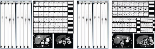
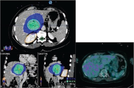
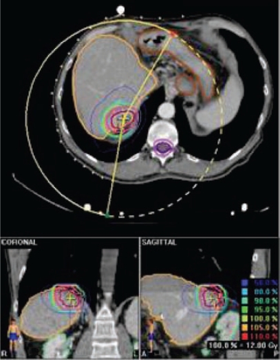
Results: Surgical, medical oncology, interventional radiology, nuclear medicine and radiation therapy approaches, their roles in management of liver tumours and ongoing research trials are presented and discussed in this article.

Conclusions: At the EIO a multi-disciplinary integrated approach to liver tumours is standard and several ongoing research projects are currently active in this field.


Figures

Figure 1:
Figure 1:
(a) wide excision of a superficial colorectal metastasis of the quadrate lobe of the liver; (b) resectional margin appearance; (c) gross pathology finding
Figure 2:
Figure 2:
(a) CT before combined systemic and intra-arterial chemotherapy—large lesion the right hepatic lobe, more than 7 cm in diameter; (b) CT after two cycles of combined chemotherapy—the lesion is smaller, with a diameter of less than 4.5 cm; (c) CT after right hepatic lobe resection: post-surgical small bile collection (arrow) and the evidence of left hepatic lobe hypertrophy
Figure 3:
Figure 3:
Chart showing relationship between responders and non-responders to neo-adjuvant chemotherapy and table comparing survival among patients undergoing resection of colorectal cancer liver metastases
Figure 4:
Figure 4:
Patient with a partial response to i.a./i.v. chemotherapy. (a) pre-treatment CT shows a huge lesion into the right lobe; (b) post-treatment CT confirms lesion reduction, detecting some calcifications within the treated lesions
Figure 5:
Figure 5:
(a) CT/PET before percutaneous thermal ablation of hepatic metastasis from colorectal cancer—FDG uptake by the lesion (arrow); (b) CT/PET performed six months later percutaneous treatment, clearly shows the absence of activity by the lesion (no FDG uptake)
Figure 6:
Figure 6:
Liver metastases from CRC in segment 7: (a) CT before treatment; (b) post-treatment CT shows a low-density area (= necrosis) where thermal ablation has been performed
Figure 7:
Figure 7:
(a) CT scan showing hepatic lesion within VI segment, before treatment; (b) Hepatic angiography after coil-embolization, shows complete occlusion of GDA (arrow); (c) CT scan after first cycle of HIAC: shrinkage of the lesion; (d) CT scan after three cycles of HIAC: further shrinkage of the target lesion. Patient affected by breast cancer metastases.
Figure 8:
Figure 8:
(a) pre-TACE CT shows a mild hypervascularity around the NET liver metastasis to be treated; (b) hepatic artery angiogram confirms the lesion, fed by the left hepatic artery; (c) post-TACE CT shows the good result after treatment, with a homogeneous Lipiodol uptake and an initial shrinkage.
Figure 9:
Figure 9:
(a) pre-TACE CT shows a huge hypervascular nodule of HCC located into the segment VI; (b) hepatic angiogram detects many arteries feeding the lesion to be treated; (c) post-TACE CT shows the intense Lipiodol uptake into the treated lesion, which is a little bit smaller after one month.
Figure 10:
Figure 10:
Preliminary SPECT scan after injection of Tc-macro-aggregated albumin in hepatic artery 99 m
Figure 11:
Figure 11:
(a) basal CT-PET performed before SIR-Spheres therapy in patient with metastases from breast cancer shows huge masses into the hepatic right lobe; (b) CT-PET two months after treatment confirmed FDG uptake reduction; (c) CT-PET three months after treatment showed a very minimal residual tumour tissue still alive
Figure 12:
Figure 12:
(a) pre-surgical CT shows a huge liver lesion; (b) CT after resection performed after embolization of right portal branch, confirms liver left lobe hypertrophy
Figure 13:
Figure 13:
Best objective response in a patient affected by liver metastases from non-functioning endocrine pancreatic carcinoma, treated with 90Y-DOTATOC. At the enrolment the patient had progressed elsewhere after trans-arterial chemo-embolisation, performed six months before (a, b and c: basal whole-body scan, anterior and posterior view, SPECT sections and CT sections, respectively; d, e and f: whole-body scan, anterior and posterior view SPECT sections and CT sections after therapy).
Figure 14:
Figure 14:
(a) stereotactic irradiation of CRC metastatis (total dose 36 Gy); (b) two-month follow-up PET/CT: no evidence of disease
Figure 15:
Figure 15:
Selective stereotactic irradiation of hepatic metastatis from CRC

Similar articles

  • Diagnosis and treatment of invasive squamous cell carcinoma of the skin: European consensus-based interdisciplinary guideline.
    Stratigos A, Garbe C, Lebbe C, Malvehy J, del Marmol V, Pehamberger H, Peris K, Becker JC, Zalaudek I, Saiag P, Middleton MR, Bastholt L, Testori A, Grob JJ; European Dermatology Forum (EDF); European Association of Dermato-Oncology (EADO); European Organization for Research and Treatment of Cancer (EORTC). Stratigos A, et al. Eur J Cancer. 2015 Sep;51(14):1989-2007. doi: 10.1016/j.ejca.2015.06.110. Epub 2015 Jul 25. Eur J Cancer. 2015. PMID: 26219687 Review.
  • The future of Cochrane Neonatal.
    Soll RF, Ovelman C, McGuire W. Soll RF, et al. Early Hum Dev. 2020 Nov;150:105191. doi: 10.1016/j.earlhumdev.2020.105191. Epub 2020 Sep 12. Early Hum Dev. 2020. PMID: 33036834
  • Diagnostics and Treatment of Thyroid Carcinoma.
    Jarząb B, Dedecjus M, Handkiewicz-Junak D, Lange D, Lewiński A, Nasierowska-Guttmejer A, Ruchała M, Słowińska-Klencka D, Nauman J, Adamczewski Z, Bagłaj M, Bałdys-Waligórska A, Barczyński M, Bednarczuk T, Cichocki A, Czarniecka A, Czepczyński R, Gawlik A, Hubalewska-Dydejczyk A, Jażdżewski K, Kamiński G, Karbownik-Lewińska M, Kos-Kudła B, Kułakowski A, Kuzdak K, Łącka K, Małecka-Tendera E, Niedziela M, Pomorski L, Sporny S, Stojcev Z, Syrenicz A, Włoch J, Krajewska J, Szpak-Ulczok S, Kalemba M, Buziak-Bereza M. Jarząb B, et al. Endokrynol Pol. 2016;67(1):74-107. doi: 10.5603/EP.2016.0011. Endokrynol Pol. 2016. PMID: 26884119
  • Six Sigma: not for the faint of heart.
    Benedetto AR. Benedetto AR. Radiol Manage. 2003 Mar-Apr;25(2):40-53. Radiol Manage. 2003. PMID: 12800564
  • Principles and guidelines for surgeons--management of symptomatic breast cancer. European Society of Surgical Oncology.
    Blichert-Toft M, Smola MG, Cataliotti L, O'Higgins N. Blichert-Toft M, et al. Eur J Surg Oncol. 1997 Apr;23(2):101-9. doi: 10.1016/s0748-7983(97)80001-7. Eur J Surg Oncol. 1997. PMID: 9158182 Review.

References

    1. El-Serag HB, Rudolph KL. Hepatocellular carcinoma: epidemiology and molecular carcinogenesis. Gastroenterology. 2007;132(7):2557–76. doi: 10.1053/j.gastro.2007.04.061. - DOI - PubMed
    1. Benson AB., 3rd Epidemiology, disease progression, and economic burden of colorectal cancer. J Manag Care Pharm. 2007;13(6 Suppl C):S5–18. - PMC - PubMed
    1. Miyazaki M, Kimura F, Shimizu H, Yoshidome H, Ohtsuka M, Kato A, et al. Surgical treatment for liver cancer. Current issues. Dig Surg. 2007;24(2):120–5. doi: 10.1159/000101899. - DOI - PubMed
    1. Rougier P, Mitry E, Barbare JC, Taieb J. Hepatocellular carcinoma (HCC): an update. Semin Oncol. 2007;34(2 Suppl 1):S12–20. doi: 10.1053/j.seminoncol.2007.01.007. - DOI - PubMed
    1. Llovet JM, Schwartz M, Mazzaferro V. Resection and liver transplantation for hepatocellular carcinoma. Semin Liver Dis. 2005;25(2):181–200. doi: 10.1055/s-2005-871198. - DOI - PubMed

LinkOut - more resources