

Neuroendocrine tumour arising inside a retro-rectal tailgut cyst: report of two cases and a review of the literature
- PMID: 22276050
- PMCID: PMC3223957
- DOI: 10.3332/ecancer.2011.201
Neuroendocrine tumour arising inside a retro-rectal tailgut cyst: report of two cases and a review of the literature
Abstract
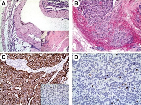
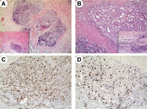
Tailgut cysts (or retro-rectal cyst-hamartomas (RCHs)) are developmental abnormalities consisting of multiloculated cysts lined by squamous, transitional or glandular epithelium which, albeit rarely, may give rise to malignant transformations. Carcinoid tumours arising in the presacral region are extremely rare and usually benign, and only a few are described in the literature. Case 1: A 63-year-old female diagnosed as having bilateral ovarian cysts underwent surgery to remove a right adnexial mass that was histopathologically diagnosed as a well-differentiated carcinoid tumour. She is currently disease free after 18 months of follow-up. Case 2: A 41-year-old-female diagnosed with hepatic metastases and a solid pelvic mass arising from a moderately differentiated neuroendocrine carcinoma is currently alive with disease after having undergone surgical removal of the mass and several medical treatments. We here describe two different clinical histories of well- and moderately differentiated neuroendocrine tumours (NETs) arising from tailgut cysts in the prerectal space together with a review of the relevant literature.
Figures
References
-
- Federspiel BH, Burke AP, Sobin LH, Shekitka KM. Rectal and colonic carcinoids. A clinicopathologic study of 84 cases. Cancer. 1990;65:135–40. - PubMed
-
- Sung MT, Ko SF, Niu CK, Hsieh CS, Huang HY. Perirenal tailgut cyst (cystic hamartoma) J Pediatr Surg. 2003;38:1404–6. - PubMed
-
- Wöhlke M, Sauer J, Dommisch K, Görling S, Hinze R. Rare case of a primary metastatic carcinoid tumour arising in a tailgut cyst. Virchows Arch. 2009;455:S390. - PubMed
Publication types
LinkOut - more resources
Full Text Sources