. 2012;7(1):e28700.
doi: 10.1371/journal.pone.0028700. Epub 2012 Jan 20.

S100B engages RAGE or bFGF/FGFR1 in myoblasts depending on its own concentration and myoblast density. Implications for muscle regeneration

Affiliations

S100B engages RAGE or bFGF/FGFR1 in myoblasts depending on its own concentration and myoblast density. Implications for muscle regeneration

Francesca Riuzzi et al. PLoS One. 2012.

Abstract

In high-density myoblast cultures S100B enhances basic fibroblast growth factor (bFGF) receptor 1 (FGFR1) signaling via binding to bFGF and blocks its canonical receptor, receptor for advanced glycation end-products (RAGE), thereby stimulating proliferation and inhibiting differentiation. Here we show that upon skeletal muscle injury S100B is released from myofibers with maximum release at day 1 post-injury in coincidence with satellite cell activation and the beginning of the myoblast proliferation phase, and declining release thereafter in coincidence with reduced myoblast proliferation and enhanced differentiation. By contrast, levels of released bFGF are remarkably low at day 1 post-injury, peak around day 5 and decline thereafter. We also show that in low-density myoblast cultures S100B binds RAGE, but not bFGF/FGFR1 thereby simultaneously stimulating proliferation via ERK1/2 and activating the myogenic program via p38 MAPK. Clearance of S100B after a 24-h treatment of low-density myoblasts results in enhanced myotube formation compared with controls as a result of increased cell numbers and activated myogenic program, whereas chronic treatment with S100B results in stimulation of proliferation and inhibition of differentiation due to a switch of the initial low-density culture to a high-density culture. However, at relatively high doses, S100B stimulates the mitogenic bFGF/FGFR1 signaling in low-density myoblasts, provided bFGF is present. We propose that S100B is a danger signal released from injured muscles that participates in skeletal muscle regeneration by activating the promyogenic RAGE or the mitogenic bFGF/FGFR1 depending on its own concentration, the absence or presence of bFGF, and myoblast density.


Conflict of interest statement

Competing Interests: The authors have declared that no competing interests exist.

Figures

Figure 1
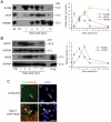
Figure 1. S100B, bFGF and HMGB1 are released from injured muscles.
(A) CM from uninjured or BaCl2-injured Tibialis anterior muscles were processed for detection of S100B, bFGF and HMGB1 by Western blotting. S100B, bFGF and HMGB1 in CM were quantified by comparing the density of individual western blot bands in the CM with that of increasing amounts of each purified protein in parallel western blots. (B) After collection of CM, muscle tissue was homogenized and subjected to Western blotting for detection of RAGE and FGFR1 (20 µg protein loaded/lane). For experiments in A and B three animals/time point were used and the whole experiment was performed two times. Results in A and B are expressed as means ± SD. C, Freshly excised uninjured or BaCl2-injured Tibialis anterior muscles were processed for detection of Pax7 and RAGE by double-immunofluorescence. Nuclei were counterstained with DAPI. RAGE is not expressed in uninjured tissue. Arrow points to a quiescent satellite (Pax7+) cell (a,a′) and to a Pax7+/RAGE+ myoblast outside regenerating myofibers (b,b′). Arrowhead points to a Pax7+/RAGE+ myoblast within a regenerating myofiber (b,b′). Bars = 100 µm.
Figure 2
Figure 2. S100B effects on differentiation vary depending on myoblast density and the duration of treatment with S100B.
(A) LD myoblasts were cultivated in DM in the absence or presence of S100B for 24 h, washed and cultivated for another 24 h in DM with no additions before MyHC immunostaining. (B) Same as in A except that LD myoblasts were subjected to Western blotting for detection of MyHC, M-cadherin and caveolin-3 at 24 h after washout (n = 3). (C) LD C2C12 myoblasts were cultivated for 1 h in DM with the p38 inhibitor, SB203580 (2 µM) or the MEK-ERK1/2 inhibitor, PD90059 (20 µM) and then for 24 h in the absence or presence of S100B, and subjected to Western blotting at 24 h after washout using anti-myogenin antibody (n = 3). (D) Same as in A except that LD myoblasts were treated with bFGF for 24 h and immunostained for MyHC at 24 and 48 h after bFGF washout. (E) LD myoblasts were cultivated in DM for 1, 24 or 72 h in the absence or presence of S100B and analyzed for ERK1/2 and p38 phosphorylation by Western blotting (n = 3). (F) LD myoblasts were cultivated in DM for 72 h in the absence or presence of S100B and subjected to MyHC immunostaining. *Significantly different from control. #Significantly different from internal control. Bars = 100 µm (A,F).
Figure 3
Figure 3. At early differentiation stages S100B exerts mitogenic effects in LD and HD myoblasts and activates the myogenic program in LD myoblasts.
(A) LD and HD myoblasts were cultivated for 24 h in DM in the absence or presence of S100B plus or minus 20 µM PD98059, 2 µM SB203580 or both before cell counts. (B) Parallel cells cultivated for 24 h in DM in the absence or presence of S100B were subjected to BrdU incorporation assay. (C) LD myoblasts were cultivated for 24 h in DM in the absence or presence of S100B and either subjected to BrdU incorporation assay or washed and cultivated in DM for another 24 or 48 h without additions before BrdU incorporation assay. Results are expressed as cell numbers/field (top panel) and percentages of BrdU+ cells (bottom panel). (D) LD myoblasts cultivated for 24 h in DM in the absence or presence of S100B plus or minus 20 µM PD98059 or 2 µM SB203580 were subjected to RT-PCR to detect myogenin mRNA. *Significantly different from control. **Significantly different from control (first column from left) and internal control.
Figure 4
Figure 4. S100B activates RAGE, but not bFGF/FGFR1 in LD myoblasts.
(A) LD myoblasts in DM were pretreated with non-immune IgG, anti-RAGE antibody or anti-FGFR1 antibody (2 h) and cultivated for 24 h in the absence or presence of S100B. Cultures were subjected to Western blotting to detect phosphorylated ERK1/2 and p38 (n = 3). (B) Same as in A except that cells pretreated with anti-RAGE or anti-FGFR1 antibody were cultivated for 24 h in the absence or presence of S100B and processed for proliferation by a BrdU incorporation assay (n = 3). (C,D) Same as in B except that cells pretreated with anti-RAGE or anti-FGFR1 antibody were cultivated for 24 h in the absence or presence of S100B, washed and cultivated in DM for another 24 h without additions and processed for MyHC immunostaining (C) or Western blotting of MyHC (D) (n = 3). *Significantly different from control (first column from left). #Significantly different from internal control. n = 3. Bars = 100 µm (C).
Figure 5
Figure 5. RAGE, but not bFGF/FGFR1 co-immunoprecipitates with S100B in LD myoblasts at early differentiation stages.
(A) LD myoblasts cultivated in DM for 30 min in the presence of 1 nM S100B were subjected to immunoprecipitation using anti-S100B, anti-FGFR1, anti-bFGF or anti-RAGE antibody. The immunoprecipitates were subjected to Western blotting for detection of S100B, FGFR1, bFGF, or RAGE. In control samples non-immune IgG (IP-IgG) were used. Twenty µg protein were loaded in input lanes. (B) LD myoblasts were cultivated in DM for 30 min in the absence or presence of either 1 nM S100B or 1 nM bFGF after pretreatment with IgG (10 µg/ml) or anti-RAGE antibody (10 µg/ml). Cells were lysed and cell lysates were subjected to Western blotting for detection of phosphorylated Tyr and FGFR1. (C) LD myoblasts cultivated in DM for 2 h in the presence of either non-immune IgG (10 µg/ml) or anti-RAGE antibody (10 µg/ml). Cultures were then added with 1 nM S100B for 30 min and subjected to immunoprecipitation using anti-S100B antibody. Immunoprecipitates were probed by Western blotting with anti-S100B, anti-FGFR1, anti-bFGF or anti-RAGE antibody. The WB:FGFR1 filter was reprobed with anti- phosphorylated Tyr antibody (marked by the “[” symbol). (D) LD myoblasts were cultivated in DM for 3 days in the presence of 1 nM S100B without medium renovation and subjected to immunoprecipitation using anti-S100B antibody. The immunoprecipitates were probed by Western blotting with anti-S100B, anti-FGFR1, anti-bFGF or anti-RAGE antibody. (E) LD myoblasts were cultivated in DM for 3 days in the presence of 1 nM S100B without medium renovation and counted. *Significantly different from control.
Figure 6
Figure 6. At high doses S100B promotes the formation of a RAGE/S100B/bFGF/FGFR1 tetracomplex in LD myoblasts at early differentiation stages.
(A) HD myoblasts (one dish) and LD myoblasts (four dishes) were cultivated in DM for 24 h. Culture media were collected and subjected to Western blotting using anti-bFGF antibody. After collection of culture media the cells were lysed for Western blotting of tubulin. (B) LD myoblasts were cultivated in DM for 30 min in the presence of 1 or 100 nM S100B and subjected to immunoprecipitation using anti-S100B antibody. The immunoprecipitates were probed with anti-FGFR1, anti-RAGE or anti-bFGF antibody. (C) LD myoblasts were cultivated in DM for 24 h in the presence of varying S100B concentrations and subjected to BrdU incorporation assay to measure proliferation at the end of the 24-h treatment (upper panel). Parallel cells were washed after the first 24-h treatment, further cultivated in DM for 24 h without additions and subjected to Western blotting for detection of MyHC (lower panel). (D) LD myoblasts were cultivated in DM for 30 min in the absence or presence of 100 nM S100B. Cells were lysed and cell lysates were subjected to Western blotting for detection of pho-Tyr and FGFR1. (E) LD myoblasts were cultivated in DM for 2 h in the presence of either 40 µg/ml non-immune IgG or 40 µg/ml anti-bFGF antibody followed by addition of vehicle or S100B to 100 nM for 30 min and immunoprecipitation using anti-S100B antibody. The immunoprecipitates were probed with anti-bFGF, anti-FGFR1 or anti-RAGE antibody. (F) LD myoblasts cultivated in DM for 30 min in the presence of 100 nM S100B were subjected to immunoprecipitation using anti-S100B, anti-FGFR1, anti-bFGF or anti-RAGE antibody. The immunoprecipitates were subjected to Western blotting for detection of S100B, FGFR1, bFGF, or RAGE. (G) LD myoblasts were cultivated in GM for 24 h, washed, scraped using PBS and collected by centrifugation. The pellet was resuspended in DM (5 ml, i.e. the same volume as used during the cultivation in petri dishes) containing 1 nM S100B, centrifuged at 500× g for 5 min at room temperature and incubated at 37°C for 30 min without aspiring the supernatant. By this procedure, cell-cell contacts of the original LD culture were augmented thus artificially reproducing the conditions of an HD culture. The supernatant was aspired, and the pellet was resuspended in PBS containing 1 mM CaCl2 and centrifuged as above. The pellet was resuspended in 1 ml of lysis buffer for subsequent immunoprecipitation using anti-S100B antibody. The immunoprecipitate was probed with anti-RAGE, anti-FGFR1, anti-S100B or anti-bFGF antibody. (H) Complex formation among S100B, bFGF, FGFR1 and RAGE in LD myoblasts as investigated by in situ proximity ligation assay (PLA). LD myoblasts in DM were incubated for 30 min with varying S100B concentrations as indicated or with 1 nM bFGF. LD myoblasts in DM were also treated with either 5 µg/ml non-immune IgG or 5 µg/ml anti-bFGF antibody followed by addition of vehicle or S100B to 100 nM for 30 min. Cells were then subjected to in situ PLA using antibody pairs as indicated on top of panels. In control experiments one antibody of individual pairs was omitted as indicated. *Significantly different from control. Bars = 20 µm (H).
Figure 7
Figure 7. S100B/RAGE and RAGE/FGFR1 complexes in regenerating skeletal muscle tissue.
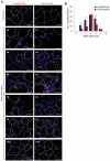
(A) Uninjured and injured skeletal muscle tissue was subjected to in situ PLA using anti-S100B/anti-RAGE antibody pairs (left) or anti-RAGE/anti-FGFR1 antibody pairs (right). Myofiber boundaries in transverse sections are marked by dotted lines. No S100B/RAGE or RAGE/FGFR1 complexes can be detected in uninjured skeletal muscle tissue. Arrows point to RAGE/FGFR1 complexes at apposed myoblasts. RAGE/FGFR1 complexes actually represent RAGE/S100B/bFGF/FGFR1 - see text). Bars = 200 µm. (B) Quantitative analysis of the distribution of S100B/RAGE and RAGE/FGFR1 complexes in uninjured skeletal muscle tissue and at the indicated days post-injury. In situ complexes were counted in 10 fields (×20 magnification)/section in a total of 10 sections/time point from 2 animals. *Significantly different.
Figure 8
Figure 8. S100B engages RAGE in LD wild-type myoblasts and activates FGFR1 in Rage −/− myoblasts.
(A) Primary myoblasts from WT and Rage −/− mice in DM were treated for 30 min with 1 nM S100B and subjected to immunoprecipitation using an anti-S100B antibody. S100B immunoprecipitates were analyzed for RAGE, FGFR1, bFGF and c-Met levels. (B) LD WT and Rage −/− myoblasts were cultivated in DM for 30 min in the absence or presence of either 1 nM S100B or 1 nM bFGF. Cells were lysed and cell lysates were subjected to Western blotting for detection of phosphorylated Tyr and FGFR1. (C) WT and Rage −/− myoblasts cultivated in DM for 24 h in the absence or presence of 1 nM S100B were subjected to BrdU incorporation assay. (D) WT and Rage −/− myoblasts were cultivated in DM in the absence or presence of 1 nM S100B for 24 or 72 h without medium renewal. One set of cells was washed and further cultivated for 48 h in DM without additions. At the end of treatments, cells were lysed and cell lysates were subjected to Western blotting to detect myogenin (as a myogenic differentiation marker). *Significantly different from control (first column from left). #Significantly different from internal control (second column from left).
Figure 9
Figure 9. Proposed schematics of S100B/RAGE and RAGE/S100B/bFGF/FGFR1 interactions in LD and HD myoblasts and of action of released S100B following skeletal muscle injury.
(A) (Left) In LD myoblast cultures S100B tetramers/octamers promote RAGE oligomerization and/or stabilization of RAGE oligomers resulting in the simultaneous stimulation of proliferation and activation of the myogenic program; this occurs irrespective of the absence or presence of bFGF. At high concentrations, high-order S100B oligomers bind bFGF and the S100B/bFGF complex cross-links RAGE and FGFR1 on the same cell. This results in enhancement of FGFR1 signaling and blockade of RAGE signaling likely due to lack of RAGE oligomerization and/or stabilization of RAGE oligomers. However, in the absence of bFGF either low or high S100B engages RAGE. (Right) In HD myoblast cultures, S100B tetramers/octamers bind bFGF and the S100B/bFGF complex cross-links RAGE and FGFR1 on apposed cells resulting in blockade of RAGE signaling likely due to lack of RAGE oligomerization and/or stabilization of RAGE oligomers and enhancement of FGFR1 signaling. This results in stimulation of proliferation and inhibition of differentiation. (B) Early after injury S100B mainly engages RAGE on activated satellite cells thereby stimulating their proliferation and activating the myogenic program. Whether S100B activates satellite cells remains to be investigated (?). During the proliferation phase S100B binds to both, RAGE and bFGF/FGFR1 and promotes the formation of a RAGE/S100B/bFGF/FGFR1 tetracomplex thereby stimulating myoblast proliferation and blocking RAGE . At this stage HMGB1 might compete with S100B for binding to RAGE thereby unlocking RAGE and stimulating its promyogenic activity . Later on, with diminishing S100B levels RAGE would be activated by HMGB1 thereby promoting myoblast differentiation and myocyte fusion into myofibers.

Similar articles

Cited by

References

    1. Chargé SB, Rudnicki MA. Cellular and molecular regulation of muscle regeneration. Physiol Rev. 2004;84:209–238. - PubMed
    1. Buckingham M. Myogenic progenitor cells and skeletal myogenesis in vertebrates. Curr Opin Genet Dev. 2006;16:525–532. - PubMed
    1. Donato R, Sorci G, Riuzzi F, Arcuri C, Bianchi R, et al. S100B's double life: intracellular regulator and extracellular signal. Biochm Biophys Acta. 2009;1793:1008–1022. - PubMed
    1. Schmidt AM, Yan SD, Yan SF, Stern DM. The multiligand receptor RAGE as a progression factor amplifying immune and inflammatory responses. J Clin Invest. 2001;108:949–955. - PMC - PubMed
    1. Bierhaus A, Humpert PM, Morcos M, Wendt T, Chavakis T, et al. Understanding RAGE, the receptor for advanced glycation end products. J Mol Med. 2005;83:876–886. - PubMed

Publication types

MeSH terms

Substances