Bone marrow-derived cells from male donors do not contribute to the endometrial side population of the recipient
- PMID: 22276168
- PMCID: PMC3261882
- DOI: 10.1371/journal.pone.0030260
Bone marrow-derived cells from male donors do not contribute to the endometrial side population of the recipient
Abstract
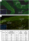
Accumulated evidence demonstrates the existence of bone marrow-derived cells origin in the endometria of women undergoing bone marrow transplantation (BMT). In these reports, cells of a bone marrow (BM) origin are able to differentiate into endometrial cells, although their contribution to endometrial regeneration is not yet clear. We have previously demonstrated the functional relevance of side population (SP) cells as the endogenous source of somatic stem cells (SSC) in the human endometrium. The present work aims to understand the presence and contribution of bone marrow-derived cells to the endometrium and the endometrial SP population of women who received BMT from male donors. Five female recipients with spontaneous or induced menstruations were selected and their endometrium was examined for the contribution of XY donor-derived cells using fluorescent in situ hybridization (FISH), telomapping and SP method investigation. We confirm the presence of XY donor-derived cells in the recipient endometrium ranging from 1.7% to 2.62%. We also identify 0.45-0.85% of the donor-derived cells in the epithelial compartment displaying CD9 marker, and 1.0-1.83% of the Vimentin-positive XY donor-derived cells in the stromal compartment. Although the percentage of endometrial SP cells decreased, possibly being due to chemotherapy applied to these patients, they were not formed by XY donor-derived cells, donor BM cells were not associated with the stem cell (SC) niches assessed by telomapping technique, and engraftment percentages were very low with no correlation between time from transplant and engraftment efficiency, suggesting random terminal differentiation. In conclusion, XY donor-derived cells of a BM origin may be considered a limited exogenous source of transdifferentiated endometrial cells rather than a cyclic source of BM donor-derived stem cells.
Conflict of interest statement
Figures


Similar articles
-
Bone marrow-derived cells from male donors can compose endometrial glands in female transplant recipients.Am J Obstet Gynecol. 2009 Dec;201(6):608.e1-8. doi: 10.1016/j.ajog.2009.07.026. Epub 2009 Oct 3. Am J Obstet Gynecol. 2009. PMID: 19800602
-
Endometrial cells derived from donor stem cells in bone marrow transplant recipients.JAMA. 2004 Jul 7;292(1):81-5. doi: 10.1001/jama.292.1.81. JAMA. 2004. PMID: 15238594
-
Experimental evidence for bone marrow as a source of nonhematopoietic endometrial stromal and epithelial compartment cells in a murine model.Biol Reprod. 2013 Jul 11;89(1):7. doi: 10.1095/biolreprod.113.107987. Print 2013 Jul. Biol Reprod. 2013. PMID: 23699390
-
Endometrial stem/progenitor cells: the first 10 years.Hum Reprod Update. 2016 Mar-Apr;22(2):137-63. doi: 10.1093/humupd/dmv051. Epub 2015 Nov 9. Hum Reprod Update. 2016. PMID: 26552890 Free PMC article. Review.
-
Somatic stem cells in the human endometrium.Semin Reprod Med. 2013 Jan;31(1):69-76. doi: 10.1055/s-0032-1331800. Epub 2013 Jan 17. Semin Reprod Med. 2013. PMID: 23329639 Review.
Cited by
-
G-CSF and stem cell therapy for the treatment of refractory thin lining in assisted reproductive technology.J Assist Reprod Genet. 2017 Jul;34(7):831-837. doi: 10.1007/s10815-017-0922-6. Epub 2017 Apr 12. J Assist Reprod Genet. 2017. PMID: 28405864 Free PMC article. Review.
-
Very small embryonic-like stem cells are the elusive mouse endometrial stem cells--a pilot study.J Ovarian Res. 2015 Mar 11;8:9. doi: 10.1186/s13048-015-0138-2. J Ovarian Res. 2015. PMID: 25824685 Free PMC article.
-
Endometrial Stem Cell Markers: Current Concepts and Unresolved Questions.Int J Mol Sci. 2018 Oct 19;19(10):3240. doi: 10.3390/ijms19103240. Int J Mol Sci. 2018. PMID: 30347708 Free PMC article. Review.
-
Uterus: A Unique Stem Cell Reservoir Able to Support Cardiac Repair via Crosstalk among Uterus, Heart, and Bone Marrow.Cells. 2022 Jul 13;11(14):2182. doi: 10.3390/cells11142182. Cells. 2022. PMID: 35883625 Free PMC article. Review.
-
Stem cell-based therapy for ameliorating intrauterine adhesion and endometrium injury.Stem Cell Res Ther. 2021 Oct 30;12(1):556. doi: 10.1186/s13287-021-02620-2. Stem Cell Res Ther. 2021. PMID: 34717746 Free PMC article. Review.
References
-
- Padykula AH. Regeneration in the primate uterus: the role of stem cells. Ann N Y Acad Sci. 1991;622:47–56. - PubMed
-
- Chan RW, Gargett CE. Identification of label- retaining cells in mouse endometrium. Stem Cells. 2006;24:1529–1538. - PubMed
-
- Cervelló I, Martinez-Conejero JA, Horcajadas JA, Pellicer A, Simón C. Identification, characterization and co-localization of label-retaining cell population in mouse endometrium with typical undifferentiated markers. Human Reproduction. 2007;22:45–51. - PubMed
-
- Cho NH, Park YK, Kim YT, Yang H, Kim SK. Lifetime expression of stem cell markers in the uterine endometrium. Fertil Steril. 2004;81(2):403–7. - PubMed
-
- Chan RW, Schwab KE, Gargett CE. Clonogenicity of human endometrial epithelial and stromal cells. Biol Reprod. 2004;70(6):1738–50. - PubMed
Publication types
MeSH terms
Substances
LinkOut - more resources
Full Text Sources
Other Literature Sources
Medical

