. 2012;7(1):e30360.
doi: 10.1371/journal.pone.0030360. Epub 2012 Jan 20.

Transforming growth factor-β1 suppresses hepatitis B virus replication by the reduction of hepatocyte nuclear factor-4α expression

Affiliations

Transforming growth factor-β1 suppresses hepatitis B virus replication by the reduction of hepatocyte nuclear factor-4α expression

Ming-Hsiang Hong et al. PLoS One. 2012.

Abstract

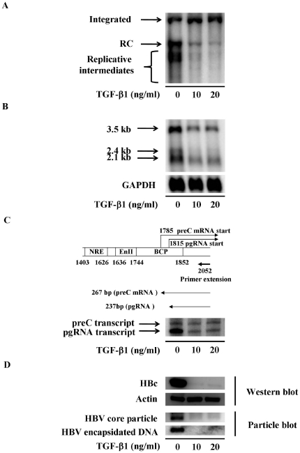
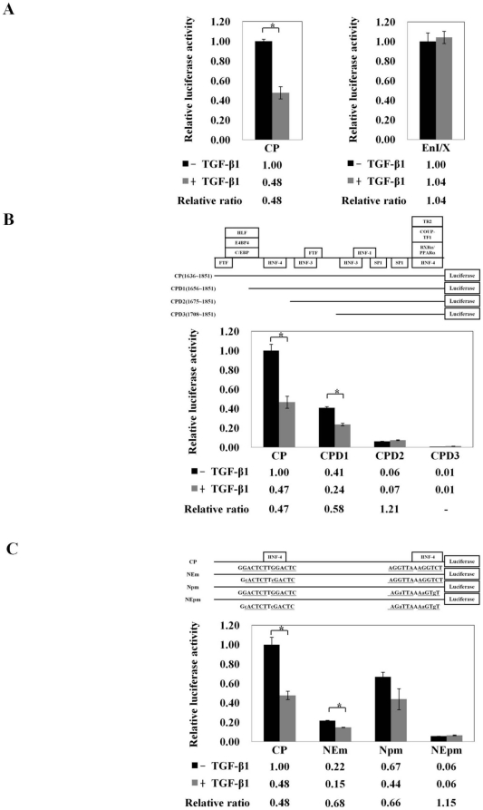
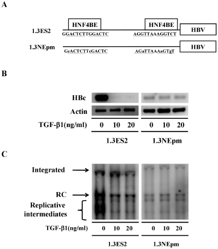
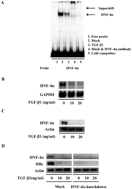
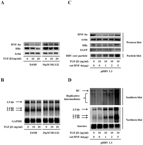
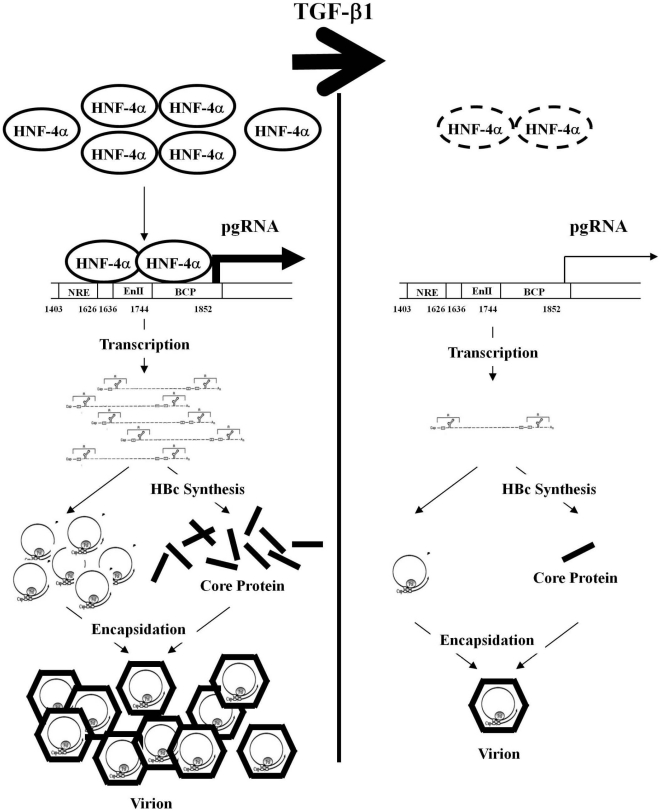
Several studies have demonstrated that cytokine-mediated noncytopathic suppression of hepatitis B virus (HBV) replication may provide an alternative therapeutic strategy for the treatment of chronic hepatitis B infection. In our previous study, we showed that transforming growth factor-beta1 (TGF-β1) could effectively suppress HBV replication at physiological concentrations. Here, we provide more evidence that TGF-β1 specifically diminishes HBV core promoter activity, which subsequently results in a reduction in the level of viral pregenomic RNA (pgRNA), core protein (HBc), nucleocapsid, and consequently suppresses HBV replication. The hepatocyte nuclear factor 4alpha (HNF-4α) binding element(s) within the HBV core promoter region was characterized to be responsive for the inhibitory effect of TGF-β1 on HBV regulation. Furthermore, we found that TGF-β1 treatment significantly repressed HNF-4α expression at both mRNA and protein levels. We demonstrated that RNAi-mediated depletion of HNF-4α was sufficient to reduce HBc synthesis as TGF-β1 did. Prevention of HNF-4α degradation by treating with proteasome inhibitor MG132 also prevented the inhibitory effect of TGF-β1. Finally, we confirmed that HBV replication could be rescued by ectopic expression of HNF-4α in TGF-β1-treated cells. Our data clarify the mechanism by which TGF-β1 suppresses HBV replication, primarily through modulating the expression of HNF-4α gene.


Conflict of interest statement

Competing Interests: The authors have declared that no competing interests exist.

Figures

Figure 1
Figure 1. TGF-β1 represses viral replication in HBV expressing cells.
1.3ES2 cells were treated with or without TGF-β1 for 6 days. (A) Total DNA was extracted followed by HindIII digestion. The viral replicative intermediates were examined by Southern blot analysis using an HBV-specific probe. The intensity of the integrated HBV genome was used as internal control for the equal amount of sample loading. Bands corresponding to the integrated genome (integrated), the relaxed circular double-stranded DNA (RC), the replicative intermediates are indicated individually. (B) Total RNA was extracted, and the expression of viral transcripts were examined by Northern blot analysis using HBV-specific probe. Expression of GAPDH was used as an internal control. (C) The amount of pgRNA and pre-C transcripts was analyzed by primer extension analysis. The schematic illustration shows the relative position of the HBV-specific primers used for primer extension analysis. (D) The cell lysate was harvested, and the expression level of HBc was determined by Western blot analysis. HBV nucleocapsids and the embedded viral gnome were detected by using particle blot analysis. Expression of actin was used as an internal control.
Figure 2
Figure 2. The HNF-4α binding sites within HBV core promoter are the essential elements for the inhibitory effects of TGF-β1.
For the promoter activity assay, HepG2 cells were transfected with luciferase reporter plasmids driven by HBV core promoter (CP) and enhancer I/X promoter (EnI/X) (A), with luciferase reporter plasmids driven by HBV core promoter (CP) and truncated HBV core promoters (CPD1, CPD2 and CPD3) (B), or with luciferase reporters with HNF4BE(s)-mutated HBV core promoters (NEm, Npm and NEpm) (C). Two days after TGF-β1 treatment, the cell lysate was extracted for luciferase activity analysis. The luciferase activities from cells transfected with different reporter plasmids were normalized to the galactosidase activities from co-transfected pCMV-beta-galactosidase plasmids, and the relative luciferase activities were compared to that of core promoter (CP) or enhancer I/X promoter (EnI/X), respectively. The relative ratio of their promoter activities between TGF-β1-treated cells and corresponding mock control cells were shown below. The schematic illustration shows the reporter constructs with deletions or specific mutations of HNF-4α binding sites in the HBV core promoter region. The results were shown as relative ratio of the firefly luciferase activities normalized to the beta-galactosidase activities in triplicates (mean value±S.D). Statistical analyses were carried out by 2-sided paired t test, and the star symbol representing the p values less than 0.01 were considered as statistically significant.
Figure 3
Figure 3. The HNF-4α binding sites within the HBV core promoter are essential elements for the TGF-β1-mediated suppression.
(A) Schematic illustration of HBV-expressing plasmids with HNF-4α binding sites mutations within the HBV core promoter region. (B) Stably HBV-producing cell lines (1.3ES2 and 1.3NEpm) were treated with or without TGF-β1 for 6 days. To determine the expression level of HBc, cell lysate was extracted and subjected to Western blot analysis. The loaded amount of total protein was adjusted by the expression level of actin. (C) Total DNA was extracted from stable HBV-producing cell lines followed by HindIII digestion, and the viral replicative intermediates were examined by Southern blot analysis using an HBV-specific probe. The integrated HBV genomes were revealed as the internal control for equal amount of sample loading. Bands corresponding to the integrated genome (integrated), the relaxed circular double-stranded DNA (RC), the replicative intermediates are indicated individually.
Figure 4
Figure 4. TGF-β1 treatment represses HNF-4α expression in HBV-producing cells.
(A) The binding activity of endogenous HNF-4α in the presence or absence of TGF-β1 was examined by EMSA analysis. Nuclear extracts were prepared from HepG2 cells with or without TGF-β1 treatment for 2 days, and then incubated with HBV-specific probes containing HNF-4α binding element. Lane 1 is a free probe control without any nuclear extracts. Lane 2 is the specific probe incubated with HepG2 nuclear extracts. Lane 3 is the specific probe incubated with TGF-β1-treated nuclear extracts. Lane 4 is the specific probe incubated with HepG2 nuclear extracts and antibody against HNF-4α. Lane 5 is the specific probe incubated with HepG2 nuclear extracts and 100-fold molar excess cold competitor. (B) 1.3ES2 cells were plated followed by TGF-β1 treatment for 6 days. Total RNA was extracted analyzed by Northern blot with an HNF-4α specific probe. The expression of GAPDH was used as loading control. (C) 1.3ES2 cells were treated with TGF-β1 for 6 days. To examine the expression of HNF-4α, the cell lysate was extracted and subjected by Western blot analysis. The loaded amount of total protein was adjusted by the expression level of actin. (D) 1.3ES2 cells were infected with lentivirus carrying HNF-4α shRNA. The transduced cells were selected with puromycin, and then cells were treated by TGF-β1 for 6 days. The cell lysate was collected for Western blot analysis to determine the expression level of HNF-4α and HBc. The total protein loaded was adjusted by actin expression level.
Figure 5
Figure 5. HNF-4α plays a crucial role in the TGF-β1-associated inhibitory effect on HBV replication.
1.3ES2 cells were treated with or without 10 µM MG132 for 1 hour and TGF-β1 for 12 hours. (A) The cell lysate was collected for Western blot analysis for the detection of HNF-4α and HBc. The loading amount of total protein was monitored by actin. (B) Total RNA was extracted, and the expression of HBV transcripts were revealed by Northern blot with using an HBV-specific probe. The expression of GAPDH was used as the loading control. (C) HepG2 cells were transfected with pHBV1.3 plasmids and an increased amount of rat HNF-4α-expressing plasmids. After treated with TGF-β1 for 2 days, the cell lysate was collected for the detection of HNF-4α and HBc by Western blot analysis. The expression of core particles was examined by particle blot analysis. The equal amount of total protein was revealed by the level of actin. The amount of HBc and HBV particle were also adjusted by the expression level of EGFP for monitoring the transfection efficiency. (D) Total RNA was extracted, and the expression of HBV transcripts were revealed by Northern blot with an HBV-specific probe. The expression of the kanamycin-neomycin phosphate transferase (kan/neo) was used as the loading control. Total DNA was extracted followed by HindIII digestion. The viral replicative intermediates were examined by Southern blot analysis using an HBV-specific probe. Bands corresponding to the relaxed circular double-stranded DNA (RC), the replicative intermediates are indicated individually.
Figure 6
Figure 6. Schematic illustration of our model in which TGF-β1 exerts its anti-HBV effect through the modulation of cellular HNF-4α expression level.
In the hepatocytes, the abundantly expressed HNF-4α efficiently activates the transcription of HBV pgRNA. The accumulated pgRNA not only serve as the templates for synthesis of viral core protein but also assemble with HBV core protein to form viral nucleocapsids. In TGF-β1-treated hepatocytes, the expression level of HNF-4α is dramatically reduced, which severely interfere with the synthesis of pgRNA and HBV core protein. Lacking of those viral components, the formation of nucleocapsid and HBV replication are substantially suppressed.

Similar articles

Cited by

References

    1. Chen CJ, Yang HI, Su J, Jen CL, You SL, et al. Risk of hepatocellular carcinoma across a biological gradient of serum hepatitis B virus DNA level. JAMA. 2006;295:65–73. - PubMed
    1. Farazi PA, DePinho RA. Hepatocellular carcinoma pathogenesis: from genes to environment. Nat Rev Cancer. 2006;6:674–687. - PubMed
    1. Seeger C, Mason WS. Hepatitis B virus biology. Microbiol Mol Biol Rev. 2000;64:51–68. - PMC - PubMed
    1. Moolla N, Kew M, Arbuthnot P. Regulatory elements of hepatitis B virus transcription. J Viral Hepat. 2002;9:323–331. - PubMed
    1. Tang H, Banks KE, Anderson AL, McLachlan A. Hepatitis B virus transcription and replication. Drug News Perspect. 2001;14:325–334. - PubMed

Publication types

MeSH terms

Substances