. 2012;7(1):e30759.
doi: 10.1371/journal.pone.0030759. Epub 2012 Jan 20.

Dedifferentiation of foetal CNS stem cells to mesendoderm-like cells through an EMT process

Affiliations

Dedifferentiation of foetal CNS stem cells to mesendoderm-like cells through an EMT process

Suzan Ber et al. PLoS One. 2012.

Abstract

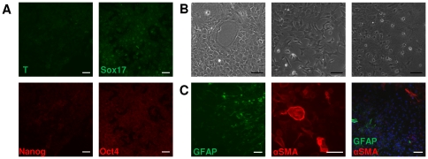
Tissue-specific stem cells are considered to have a limited differentiation potential. Recently, this notion was challenged by reports that showed a broader differentiation potential of neural stem cells, in vitro and in vivo, although the molecular mechanisms that regulate plasticity of neural stem cells are unknown. Here, we report that neural stem cells derived from mouse embryonic cortex respond to Lif and serum in vitro and undergo epithelial to mesenchymal transition (EMT)-mediated dedifferentiation process within 48 h, together with transient upregulation of pluripotency markers and, more notably, upregulation of mesendoderm genes, Brachyury (T) and Sox17. These induced putative mesendoderm cells were injected into early gastrulating chick embryos, which revealed that they integrated more efficiently into mesoderm and endoderm lineages compared to non-induced cells. We also found that TGFβ and Jak/Stat pathways are necessary but not sufficient for the induction of mesendodermal phenotype in neural stem cells. These results provide insights into the regulation of plasticity of neural stem cells through EMT. Dissecting the regulatory pathways involved in these processes may help to gain control over cell fate decisions.


Conflict of interest statement

Competing Interests: The work was supported by The Technology Programme (DTI Project Number TP/4/BIO/6/I/22020) of CellCentric Ltd. This does not alter the authors' adherence to all the PLoS ONE policies on sharing data and materials.

Figures

Figure 1
Figure 1. Expression of pluripotency associated proteins in serum-free cultured neurospheres and 48 h serum and Lif induced neurospheres.
(A): Morphology in serum-free (upper panel) and 48 h serum/Lif (lower panel) conditions. (B): Transcriptional profile of pluripotency associated factors Oct4, Sox2, c-Myc, Klf4 and Nanog of serum-free cultured neurospheres and 48 h treated neurospheres. Data is expressed as ΔΔCT and normalized to ES cells. (C): Immunostaining for Oct4, Nanog, Sox2, SSEA1 and alkaline phosphatase. Abbreviations: AP, alkaline phosphatase; SSEA1, stage specific embryonic antigen 1. Scale bars: 50 µm.
Figure 2
Figure 2. EMT and mesendoderm markers upregulated in neurospheres after 48 h induction with serum and Lif.
(A): Immunostaining for Slug, E-cadherins and N-cadherins (B): Immunostaining for mesendoderm markers Sox17 and Brachyury (T). (C): Expression profile of Slug, N-cadherins, E-cadherins, Sox17 and Brachyury of 48 h serum and Lif induced neurospheres measured by QPCR relative to neurospheres cultured in standard neural stem cell media. Abbreviations: Ncad, N-cadherins; Ecad, E-cadherins. Scale bars: 50 µm.
Figure 3
Figure 3. In vitro differentiation of neurospheres in serum and Lif conditions.
(A): Immunostaining for Brachyury (T), Sox17, Nanog and Oct4 at 5 days after induction in serum and Lif conditions (B): At 10 days post induction with serum and Lif, cells exhibit very heterogeneous morphologies, indicating the presence of different cell types. (C): Immunostaining for GFAP and αSMA in 10 day induced cultures. Abbreviations: GFAP, glial fibrillary acidic protein; αSMA, alpha smooth muscle actin. Scale bars: 50 µm.
Figure 4
Figure 4. In vivo integration potential of 48 h serum and Lif induced neurospheres.
(A): Diagram showing injection site of induced (green) and non-induced (red) neurospheres in HH3+ chick embryos. B,D show images taken immediately after injection, and 24 h (stage HH8-9) post injection of two different embryos. Cross section images correspond to planes indicated on representative images of fixed embryos 24 h after injection. (B): Cross sections show successful integration of injected cells in the lateral plate mesoderm and endoderm (1, 2). (C): both green and red labelled cells are detected in notochord and somite mesoderm (1). However, primarily green labelled cells have been detected ingressing from the late primitive streak (2). D, E and F show images taken immediately after injection and 40 h (stage HH12) post-injection of three different embryos. Cross section images correspond to planes indicated on representative images of fixed embryos 40 h after injection. (D): Cross section images showing injected cells within the neural tube (1) and hindbrain region (2). (E): Cross section images showing the incorporation of injected cells into sinoatrial region of the embryo (1, 2). (F): Cross sections show successful integration of injected cells in endoderm as well as lateral plate mesoderm (1, 2). (G): The chart shows proportion of green (48 h induced) and red (non-induced) cells in endoderm, mesoderm and ectoderm layers. 48 h induced cells (grey part) represent around 80% of labelled cells which integrated to mesoderm and ectoderm layers, and around 55% of labelled cells present in the ectoderm. Abreviations: HH stage, Hamburger and Hamilton stage; NC, notochord; NT, neural tube; PS, primitive streak; NP, neural plate; Endo, endoderm; Meso, mesoderm; SA, sinoatrial region.
Figure 5:
Figure 5:. In vivo differentiation of 48 h induced neural stem cells.
Staining of chick embryo paraffin sections for mesenchymal marker N-cadherin, tight junction marker ZO-1, endoderm marker Sox17, and epithelial marker E-cadherin. Although some of the labelled cells express E-cadherin, the staining pattern suggest the cells do not achieve a complete integration to ectoderm lineage. However, high integration towards mesoderm and endoderm lineages is confirmed by N-cadherin, ZO-1 and Sox17 stainings. Although induced (green) cells show higher efficiency of integration, both induced (green) and non-induced (red) cells once incorporated into these tissues express respective lineage markers and acquire similar morphological characteristics to their neighbouring host cells.
Figure 6
Figure 6. Effect of inhibitors on the expression of mesendoderm markers and morphology of neurospheres.
Jak I Inhibitor, Mek inhibitor (PD0325901), TGFβ inhibitors (Noggin+ SB431542) have been used alone or in combination as indicated. The combination of Jak I and TGFβ inhibitors resulted in the most striking inhibition of Brachyury (T) and Sox17 upregulation in neural stem cells. T (green), Sox17 (red). See result section for details. Scale bars: 50 µm.
Figure 7
Figure 7. Schematic diagram depicting our interpretation of the results.
Neural stem cells derived from later stages of development have been shown to reprogram to iPS cells via overexpression of Oct4 transcription factor (Kim et al., 2009). Signalling alone (BMPs and bFGF), however, can induce dedifferentiation of neural stem cells into a neural crest phenotype (Sailer et al., 2005). TGFβ and Jak/Stat pathways can induce a further dedifferentiation to mesendoderm-like phenotype providing evidence for extracellular signaling regulated cell plasticity of neural stem cells (this work, red arrow).

References

    1. Gilbert SF. Developmental Biology. 2006.
    1. Heisenberg CP, Solnica-Krezel L. Back and forth between cell fate specification and movement during vertebrate gastrulation. Curr Opin Genet Dev. 2008;18:311–316. - PMC - PubMed
    1. Thiery JP, Acloque H, Huang RY, Nieto MA. Epithelial-mesenchymal transitions in development and disease. Cell. 2009;139:871–890. - PubMed
    1. Acloque H, Adams MS, Fishwick K, Bronner-Fraser M, Nieto MA. Epithelial-mesenchymal transitions: the importance of changing cell state in development and disease. Journal of Clinical Investigation. 2009;119:1438–1449. - PMC - PubMed
    1. Janda E, Lehmann K, Killisch I, Jechlinger M, Herzig M, et al. Ras and TGF[beta] cooperatively regulate epithelial cell plasticity and metastasis: dissection of Ras signaling pathways. J Cell Biol. 2002;156:299–313. - PMC - PubMed

Publication types

MeSH terms