

Cerebrospinal fluid rhinorrhea: An institutional perspective from Pakistan
- PMID: 22276229
- PMCID: PMC3262998
- DOI: 10.4103/2152-7806.90689
Cerebrospinal fluid rhinorrhea: An institutional perspective from Pakistan
Abstract
Background: The management of cerebrospinal fluid (CSF) rhinorrhea has evolved over the last two decades. We present here a review of our 11-year data on CSF rhinorrhea and its management at a tertiary care hospital in a developing country, with particular reference to the diagnosis, surgical management and outcome of the disease.
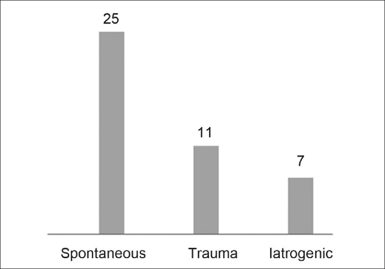
Methods: The medical charts of all patients with a diagnosis of CSF rhinorrhea over an 11-year period were reviewed. The etiology of CSF rhinorrhea was classified into three categories: spontaneous, iatrogenic and traumatic. All the patients were divided into three categories based on the type of management as conservative, intracranial and transnasal endoscopic groups.
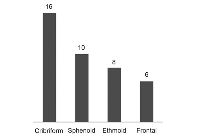
Results: A total of 43 patients fulfilled our inclusion criteria and were included in the final analysis. Eleven of the 43 patients were managed conservatively, while 22 underwent intracranial repairs; 10 patients had transnasal endoscopic repairs. The primary success rate for the transnasal approach was 70% compared to 86% for the intracranial repair. Blood loss, special care unit (SCU) stay and total cost were found to be significantly less in the transnasal endoscopic group. Computed tomography (CT) cisternography was found to have the highest sensitivity and specificity. Further, no postoperative complications were found in the transnasal endoscopic group, while five patients from the intracranial group developed various complications.
Conclusions: We conclude that the transnasal endoscopic approach has comparable success rates with the intracranial approach and significantly lower morbidity.
Keywords: Cerebrospinal fluid rhinorrhea/diagnosis; cerebrospinal fluid rhinorrhea/etiology; cerebrospinal fluid rhinorrhea/surgery; endoscopy; retrospective studies; treatment outcome.
Figures
Similar articles
-
[Clinical analysis of transnasal endoscopic repair of cerebrospinal fluid rhinorrhea].Lin Chuang Er Bi Yan Hou Tou Jing Wai Ke Za Zhi. 2019 Dec;33(12):1189-1195;1199. doi: 10.13201/j.issn.1001-1781.2019.12.018. Lin Chuang Er Bi Yan Hou Tou Jing Wai Ke Za Zhi. 2019. PMID: 31914272 Chinese.
-
Retrospective Analysis of Transnasal Endoscopic CSF Rhinorrhea Repair in a Tertiary Care Centre.Indian J Otolaryngol Head Neck Surg. 2022 Oct;74(Suppl 2):1328-1333. doi: 10.1007/s12070-021-02462-7. Epub 2021 Feb 25. Indian J Otolaryngol Head Neck Surg. 2022. PMID: 36452772 Free PMC article.
-
[Analysis of intracranial infection factors after transnasal endoscopic repair of cerebrospinal fluid rhinorrhea].Zhonghua Er Bi Yan Hou Tou Jing Wai Ke Za Zhi. 2014 Feb;49(2):121-4. Zhonghua Er Bi Yan Hou Tou Jing Wai Ke Za Zhi. 2014. PMID: 24742510 Chinese.
-
Endoscopic endonasal CSF rhinorrhea repair in children: Systematic review with meta-analysis.Int J Pediatr Otorhinolaryngol. 2020 Jul;134:110044. doi: 10.1016/j.ijporl.2020.110044. Epub 2020 Apr 10. Int J Pediatr Otorhinolaryngol. 2020. PMID: 32320837
-
Endoscopic repair of cerebrospinal fluid rhinorrhoea.Eur Ann Otorhinolaryngol Head Neck Dis. 2016 Jun;133(3):187-90. doi: 10.1016/j.anorl.2015.05.010. Epub 2016 Jan 6. Eur Ann Otorhinolaryngol Head Neck Dis. 2016. PMID: 26776882 Review.
Cited by
-
Endoscopic management of cerebrospinal fluid rhinorrhea.Asian J Neurosurg. 2016 Jul-Sep;11(3):183-93. doi: 10.4103/1793-5482.145101. Asian J Neurosurg. 2016. PMID: 27366243 Free PMC article. Review.
-
Topical Intranasal Fluorescein to Diagnose and Localize Cerebrospinal Fluid Leak: A Systematic Review.Turk Arch Otorhinolaryngol. 2021 Sep;59(3):223-229. doi: 10.4274/tao.2021.2021-3-12. Epub 2021 Oct 15. Turk Arch Otorhinolaryngol. 2021. PMID: 34713008 Free PMC article.
-
Endoscopic Surgical Repair of a Giant, Postoperative, Neglected Meningoencephalocele.Cureus. 2020 Jan 22;12(1):e6739. doi: 10.7759/cureus.6739. Cureus. 2020. PMID: 32133261 Free PMC article.
-
Application of a three-dimensional printed model to localize a cranial cerebrospinal fluid leak: a case report.J Int Med Res. 2022 Feb;50(2):3000605221078412. doi: 10.1177/03000605221078412. J Int Med Res. 2022. PMID: 35220787 Free PMC article.
-
High-Resolution Computed Tomography as an Initial Diagnostic and Localization Tool in Patients with Cerebrospinal Fluid Rhinorrhea: A Meta-Analysis.Medicina (Kaunas). 2023 Mar 10;59(3):540. doi: 10.3390/medicina59030540. Medicina (Kaunas). 2023. PMID: 36984541 Free PMC article. Review.
References
-
- Dandy WD. Pneumocephalus(intracranial pneumatocele or aerocele) Arch Surg. 1926;12:949–82.
-
- Dohlman G. Spontaneous cerebrospinal fluid rhinorrhea. Acta Otolaryngol Suppl (Stockh) 1948;67:20–3. - PubMed
-
- Giddings NA, Brackmann DE. Surgical treatment of difficult cerebrospinal fluid otorhinorrhea. Am J Otol. 1994;15:781–4. - PubMed
-
- Gulcek SY, Erdogan A, Toprak U, Ortac AA, Pasaoglu E. [Evaluation of rhinorrhea by computed tomography cisternography] Tani Girisim Radyol. 2003;9:327–32. - PubMed
-
- Har-El G. What is “spontaneous” cerebrospinal fluid rhinorrhea. Classification of cerebrospinal fluid leaks? Ann Otol Rhinol Laryngol. 1999;108:323–6. - PubMed
LinkOut - more resources
Full Text Sources
