

Recurrence of a cerebral arteriovenous malformation following complete surgical resection: A case report and review of the literature
- PMID: 22276230
- PMCID: PMC3262997
- DOI: 10.4103/2152-7806.90692
Recurrence of a cerebral arteriovenous malformation following complete surgical resection: A case report and review of the literature
Abstract
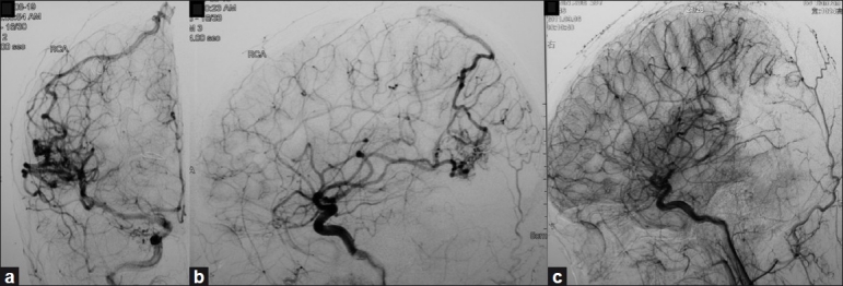
Background: Angiography-confirmed complete resection of an arteriovenous malformation (AVM) has traditionally been considered curative. However, recurrence of AVM following angiographically proven complete resection does exist, especially in children. This rare occurrence has been reported 29 times in the English language literature. Although recurrence may be asymptomatic, many reported cases result in epilepsy or intracranial hemorrhage anywhere from 0.5 to 9 years following complete resection. We report a rare case of AVM recurrence that became symptomatic 16 years after complete resection. We review the literature and discuss the relevance of performing follow-up imaging to detect AVM recurrence.
Case description: An 8-year-old girl presented with a right occipital hemorrhage with intraventricular extension from a ruptured AVM of the right occipital lobe. She underwent AVM resection through a right occipital craniotomy. Postoperative angiography confirmed complete resection and she made an uneventful recovery. Sixteen years later, she presented with a 2-month history of headaches, nausea and dizziness. Angiography revealed recurrence of the AVM which was completely resected, as documented on postoperative angiography.
Conclusion: In children, an AVM may recur after angiography-proven complete resection. Recurrence may be due to persistence and growth of an initially angiographically occult arteriovenous shunt left in place during surgery or the development of a new AVM. In addition to obtaining follow-up angiography 6-12 months after surgery, a late angiography 5 years after resection may be warranted in patients at risk for recurrence. Asymptomatic recurrence detection allows treatment and may prevent the morbidity associated with intracranial hemorrhage.
Keywords: Arteriovenous malformation; cerebral hemorrhage; postoperative angiography; recurrence.
Figures


Similar articles
-
Reappearance of arteriovenous malformations after complete resection of ruptured arteriovenous malformations: true recurrence or false-negative early postoperative imaging result?J Neurosurg. 2017 Apr;126(4):1088-1093. doi: 10.3171/2016.3.JNS152846. Epub 2016 May 27. J Neurosurg. 2017. PMID: 27231973
-
An adult case of recurrent arteriovenous malformation after "complete" surgical excision: a case report.Surg Neurol. 1999 Aug;52(2):156-8; discussion 158-9. doi: 10.1016/s0090-3019(99)00060-9. Surg Neurol. 1999. PMID: 10447283
-
Ruptured tectal arteriovenous malformation demonstrated angiographically after removal of an unruptured occipital lobe arteriovenous malformation.Neurol Med Chir (Tokyo). 2009 Jan;49(1):30-2. doi: 10.2176/nmc.49.30. Neurol Med Chir (Tokyo). 2009. PMID: 19169000
-
Resection of a posterior fossa arteriovenous malformation complicated by leaked Onyx: a case report and review of literature.Acta Neurochir (Wien). 2020 Apr;162(4):923-928. doi: 10.1007/s00701-019-04199-3. Epub 2020 Jan 30. Acta Neurochir (Wien). 2020. PMID: 31997070 Review.
-
[Coexistence of cerebral aneurysm and angiographically occult AVM in the occipital lobe; a case report].No Shinkei Geka. 1992 Mar;20(3):267-71. No Shinkei Geka. 1992. PMID: 1557177 Review. Japanese.
Cited by
-
Recurrence of a paediatric arteriovenous malformation 9 years postcomplete excision: case report and review of literature.BMJ Case Rep. 2012 Sep 25;2012:bcr2012006826. doi: 10.1136/bcr-2012-006826. BMJ Case Rep. 2012. PMID: 23010462 Free PMC article. Review.
-
Long-term outcomes in pediatric unruptured brain arteriovenous malformation treated by nonconservative management: a single center analysis.Childs Nerv Syst. 2019 Aug;35(8):1363-1369. doi: 10.1007/s00381-019-04221-0. Epub 2019 Jun 15. Childs Nerv Syst. 2019. PMID: 31201498
-
Ruptured Arteriovenous Malformation Anterior to the Brainstem to a Child with Subsequent Spontaneous Thrombosis: Case Report and Literature Review.Am J Case Rep. 2020 May 1;21:e923289. doi: 10.12659/AJCR.923289. Am J Case Rep. 2020. PMID: 32355154 Free PMC article. Review.
-
Cerebral Arteriovenous Malformation Recurrence After Complete Surgical Excision in an Adult: Case Report and Review of the Literature.Cureus. 2021 Jun 1;13(6):e15366. doi: 10.7759/cureus.15366. eCollection 2021 Jun. Cureus. 2021. PMID: 34249522 Free PMC article.
-
Recurrent arteriovenous malformation on palate after embolization combined surgical resection: preoperative magnetic resonance features and intraoperative angiographic findings.J Korean Assoc Oral Maxillofac Surg. 2015 Dec;41(6):346-51. doi: 10.5125/jkaoms.2015.41.6.346. Epub 2015 Dec 17. J Korean Assoc Oral Maxillofac Surg. 2015. PMID: 26734564 Free PMC article.
References
-
- Abdulrauf SI, Malik GM, Awad IA. Spontaneous angiographic obliteration of cerebral arteriovenous maformations. Neurosurgery. 1999;44:280–8. - PubMed
-
- Ali MJ, Bendok BR, Rosenblatt S, Rose JE, Getch CC, Batjer HH. Recurrence of pediatric cerebral arteriovenous malformations after angiographically documented resection. Pediatr Neurosurg. 2003;39:32–8. - PubMed
-
- Amacher AL, Allcock JM, Drake CG. Cerebral angiomas: The sequelae of surgical treatment. J Neurosurg. 1971;37:571–5. - PubMed
-
- Andaluz N, Myseros JS, Sathi S, Crone KR, Tew JM., Jr Recurrence of cerebral arteriovenous malformations in children: Report of two cases and review of the literature. Surg Neurol. 2004;62:324–30. - PubMed
-
- Cobb PJ, Mitha AP, Ogilvy CS. A recurrent cerebral arteriovenous malformation in an adult: Case report. J Neurosurg. 2008;109:486–91. - PubMed
Publication types
LinkOut - more resources
Full Text Sources
