

Telecytology: Clinical applications, current challenges, and future benefits
- PMID: 22276242
- PMCID: PMC3263027
- DOI: 10.4103/2153-3539.91129
Telecytology: Clinical applications, current challenges, and future benefits
Abstract
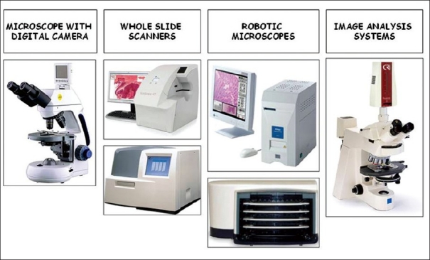
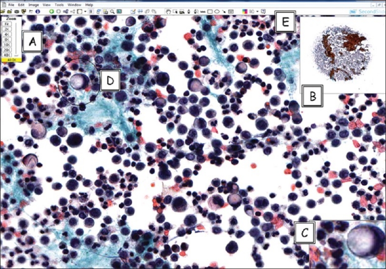
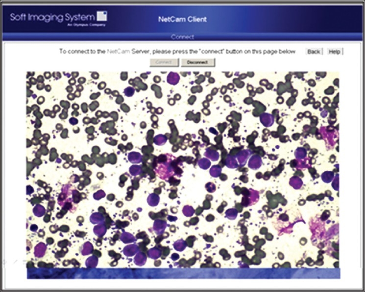
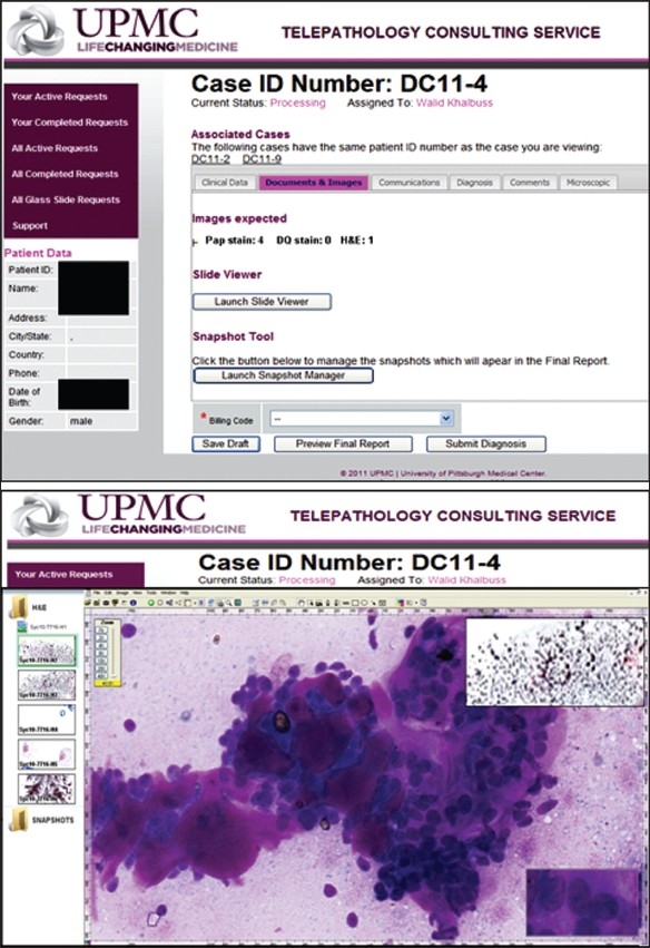
Telecytology is the interpretation of cytology material at a distance using digital images. For more than a decade, pioneering efforts to introduce telecytology into clinical practice have been reported. A Medline search for "telecytology" and "cytology" reveals a voluminous literature, though much of what has been published to date is based on technologies that are rapidly becoming obsolete. The technological limitations of previous techniques, including the transmission of static digital images and dynamic streaming images, have limited telecytology to minor niches. The primary problem with these technologies is that the remote viewer can only see a small fraction of the material on the original slides, introducing the possibility of diagnostic error based not only on image quality but also on image selection. Remote robotic microscopy offers one possible solution to this problem, but to date has found limited acceptance, principally attributable to slow operating times. Whole slide imaging seems to be a much more promising solution, though cytology-specific literature regarding its use is still scant. The advent of whole slide imaging opens up new possibilities for telecytology by enabling high-quality images of entire cytology specimens to be available to anyone, anywhere via the Internet. Although challenges remain, especially with regard to capturing the full microscopy experience including multiple planes of focus and sharp high-powered images, rapidly advancing technology promises to overcome these limitations. Increasing application of whole slide imaging technology in surgical pathology will undoubtedly also increase its application to cytology due to the increasing affordability and practicality of the equipment as it serves a larger number of useful roles within a pathology department. The current and expanding applications of telecytology for clinical practice, education, quality assurance, and testing will be reviewed.
Keywords: Digital images; remote microscopy; telecytology; whole-slide imaging.
Figures
References
-
- Yamashiro K, Shinohara T, Mitsuhashi T, Sugimura T, Taira K, Azuma M, et al. Z-axis video for cytology database is a useful tool for the case presentation prior to the cytology training workshop. Diagn Cytopathol. 2011 [In Press] - PubMed
-
- Hedvat CV. Digital microscopy: past, present, and future. Arch Pathol Lab Med. 2010;134:1666–70. - PubMed
-
- Jara-Lazaro AR, Thamboo TP, Teh M, Tan PH. Digital pathology: exploring its applications in diagnostic surgical pathology practice. Pathology. 2010;42:512–8. - PubMed
-
- Wilbur DC. Digital cytology: current state of the art and prospects for the future. Acta Cytol. 2011;55:227–38. - PubMed
LinkOut - more resources
Full Text Sources
Other Literature Sources
